Schematic overview of the cellular and molecular mechanisms involved in the cancer progression, including the proposed cellular and molecular mechanisms in cancer cells trajectory. AT1: alveolar type 1 cells; AT2: alveolar type 2 cells; AAH: atypical adenomatous hyperplasia; AIS: adenocarcinoma in situ; MIA: minimally invasive adenocarcinoma; IA: invasive adenocarcinoma; EMT: epithelial-mesenchymal transition

Home

Download

Statistics

Help

Contact

Center for Computational Systems Medicine
leaf

Tissue samples summary

leaf

Annotation details for different cell compartments

leaf

Cell type statistics by disease state

leaf

Cell type dynamics in malignant transformation

leaf

Cell lineage trajectory inference

leaf

TF regulatory network analysis

leaf

Cell-cell interaction analysis

leaf

Stemness, Senescence, Metaplasia and NeoTCR signature analysis

Tissue: Breast

Tissue samples summary

check button Cell type statistics by tissue samples.
EpitheliaImmuneStromal
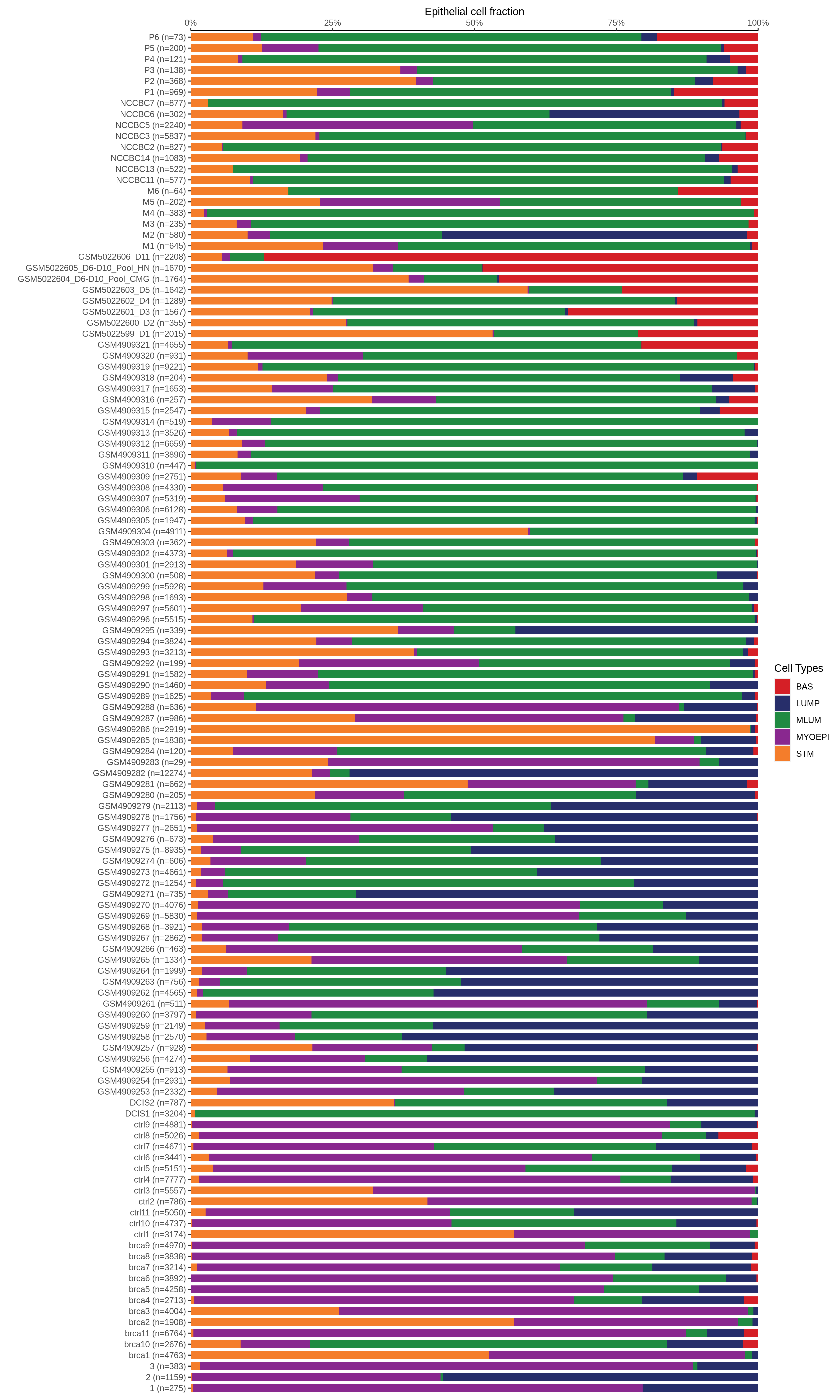
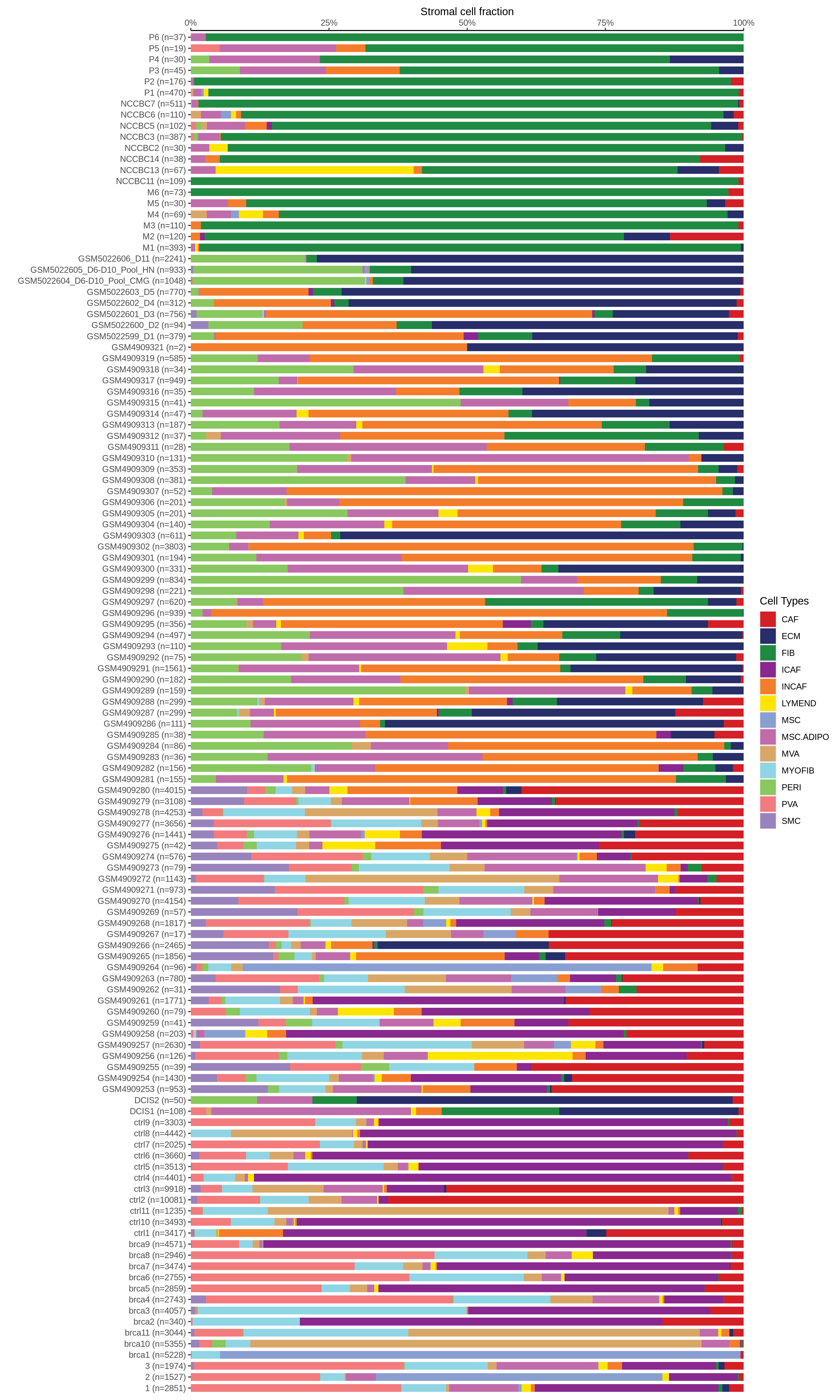
Bar plot representation of the fraction of all Epithelial cells in each sample composed of each cell type for the integrated scRNA-seq datasets of Breast tissues. Each row represents a single sample, with each color representing a different cell type present in the sample.Bar plot representation of the fraction of all Immune cells in each sample composed of each cell type for the integrated scRNA-seq datasets of Breast tissues. Each row represents a single sample, with each color representing a different cell type present in the sample.Bar plot representation of the fraction of all Stromal cells in each sample composed of each cell type for the integrated scRNA-seq datasets of Breast tissues. Each row represents a single sample, with each color representing a different cell type present in the sample.
∗Bar plot representation of the fraction of all Epithelial, Immune and Stromal cells in each sample composed of each cell type for the integrated scRNA-seq datasets of Breast tissue. Each row represents a single sample, with each color representing a different cell type present in the sample.

check button Tissue summary table.
TissueDisease StateReplicatesEpithelia UMAPPMIDSourcePlatform
BreastBRCA1-mutGSM4909277Projection

33950524

GSE161529

10X Genomics
BreastBRCA1-mutGSM4909278Projection

33950524

GSE161529

10X Genomics
BreastBRCA1-mutGSM4909279Projection

33950524

GSE161529

10X Genomics
BreastBRCA1-mutGSM4909280Projection

33950524

GSE161529

10X Genomics
BreastIDCGSM4909281Projection

33950524

GSE161529

10X Genomics
BreastIDCGSM4909282Projection

33950524

GSE161529

10X Genomics
BreastIDCGSM4909283Projection

33950524

GSE161529

10X Genomics
BreastIDCGSM4909284Projection

33950524

GSE161529

10X Genomics
BreastIDCGSM4909285Projection

33950524

GSE161529

10X Genomics
BreastIDCGSM4909286Projection

33950524

GSE161529

10X Genomics
BreastIDCGSM4909287Projection

33950524

GSE161529

10X Genomics
BreastIDCGSM4909288Projection

33950524

GSE161529

10X Genomics
BreastIDCGSM4909289Projection

33950524

GSE161529

10X Genomics
BreastIDCGSM4909290Projection

33950524

GSE161529

10X Genomics
BreastIDCGSM4909291Projection

33950524

GSE161529

10X Genomics
BreastIDCGSM4909292Projection

33950524

GSE161529

10X Genomics
BreastIDCGSM4909293Projection

33950524

GSE161529

10X Genomics
BreastIDCGSM4909294Projection

33950524

GSE161529

10X Genomics
BreastIDCGSM4909295Projection

33950524

GSE161529

10X Genomics
BreastIDCGSM4909296Projection

33950524

GSE161529

10X Genomics
∗Projection of epithelial scRNA-seq cells from Breast diseased tissue samples into the manifold of normal tissue epithelial cells. Projected cells are colored by nearest normal cells in the projection and normal epithelial cells are colored gray.
Page: 1 2 3 4 5 6 7 

Top

Annotation details for different cell compartments

check button Single-cell atlas of expresion.
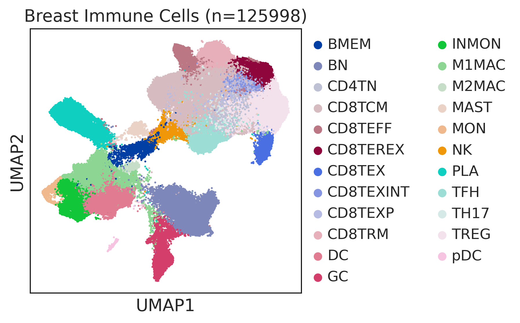
UMAP representations and annotations of Epithelial cells in the integrated scRNA-seq data of the selected tissue.UMAP representations and annotations of Immune cells in the integrated scRNA-seq data of the selected tissue.UMAP representations and annotations of Stromal cells in the integrated scRNA-seq data of the selected tissue.
∗UMAP representations and annotations of Epithelial, Immune, Stromal cells in the integrated scRNA-seq data of Breast tissue. Colors represent cell types of each compartment.

check button Cell markers.
TissueMajor Cell TypeMinor Cell TypeFull NameMarkers
BreastEpiSTMStem-like cellsALDH1A3,ALDH1A1,CD44,ITGA6,CD24,PROM1,NOTCH3,SOX9,NANOG,BMI1,POU5F1,LGR5,PROCR
BreastEpiBASBasal cellsKRT5,KRT14,SNAI2,ITGA6
BreastEpiMYOEPIMyoepithelial cellsKRT5,KRT14,TP63,ACTA2,MYLK
BreastEpiLUMPLuminal progenitorsKRT18,KRT19,LALBA,CSN2,TNFRSF11A,KIT,SLPI,PROM1,ANXA1,FOXA1,LTF,MUC1,ALDH1A3
BreastEpiMLUMMature luminal cellsESR1,PGR,FOXA1,ANKRD30A,SYTL2,GATA3,KRT8,KRT18,KRT19
BreastImmMASTMast cellsTPSAB1,CPA3,HDC,CTSG,TPSB2,CMA1,MS4A2
BreastImmDCDendritic cellsHLA-DQA1,HLA-DQB1,CLEC10A,CLEC9A,FCER1A,CST3,CD1C
BreastImmpDCPlasmacytoid dendritic cellsIL3RA,CLEC4C,NRP1,KLRD1
BreastImmNKNatural killer cellsKLRF1,SH2D1B,NCAM1,FCGR3A,GNLY,NKG7,GZMH,FGFBP2,CX3CR1,NCR1,NCR3,B3GAT1
BreastImmMONMonocytesCD14,CLEC9A,FCGR1A,LILRB2,CD209,CD1E,FCN1,VCAN
BreastImmM1MACM1 macrophagesFOLR2,FABP3,PLA2G2D,ITGAM,ITGAX,CSF1R,CD68,CD163,THBD
BreastImmINMONInflamotory monocytesCD14,CLEC9A,FCGR1A,LILRB2,CD209,CD1E,FCN1,VCAN,S100A8,S100A9
BreastImmM2MACM2 macrophagesCSF1R,CSF3R,MRC1,IL10,CCL18,VSIG4,CHI3L1
BreastImmBNNaive B cellsCD19,IGHD,IGLL1,CD27,CD38
BreastImmBMEMMemory B cellsPAX5,MS4A1,CD19,IGLL5,VPREB3,CD79A,CD79B,IGKC,CD74,HLA-DRA,CD37,CD22
BreastImmGCGerminal center B cellsSERPINA9,HRK,HTR3A,BCL6,CD180,FCRLA
BreastImmPLAPlasma cellsSSR4,IGLL5,IGLL1,AMPD1,IGHA1,IGHA2,JCHAIN,CD38,TNFRSF17,SDC1,IGHG1,MZB1
BreastImmGDTGamma-delta T cellsTRDC,TRGC1,TRGC2,NKG7,TIGIT
BreastImmCD8TEXExhausted CD8+ T cellsPDCD1,LAG3,CD101,CD38,CXCR6,TIGIT
BreastImmCD4TNNaive CD4+ T cellsIL7R,SELL,CCR7,S100A4,TCF7
BreastImmTFHT follicular helperCXCR5,BCL6,ICA1,TOX,TOX2,IL6ST,MAGEH1,BTLA,ICOS,PDCD1,CD200
BreastImmTREGRegulatory T cellsBATF,TNFRSF4,FOXP3,CTLA4,LAIR2,IL2RA
BreastImmCD8TEXPProgenitor exhausted CD8+ T cellsPDCD1,IL7R,GPR183,NR4A3,REL,TCF7
BreastImmCD8TEXINTIntermediate exhausted CD8+ T cellsPDCD1,LAG3,CD101,CD38,CXCR6,TIGIT
BreastImmCD8TEREXTerminally exhausted CD8+ T cellsTOX,GZMB,ENTPD1,ITGAE,HAVCR2,CXCL13,PDCD1,LAYN,TOX,IFNG,GZMB,MIR155HG,TNFRSF9,ITGAE
BreastImmCD8TCMCentral memory CD8+ T cellsCCR7,SELL,IL7R,CD27,CD28,PRF1,GZMA,CCL5,GPR183,S1PR1
BreastImmCD8TRMCD8+ tissue resident memory T cellsCD6,XCL1,XCL2,MYADM,CAPG,RORA,NR4A1,NR4A2,NR4A3,CD69,ITGAE
BreastImmCD8TEFFEffector CD8+ T cellsCX3CR1,FCGR3A,FGFBP2,PRF1,GZMH,TBX21,EOMES,S1PR1
BreastStrENDEndothelial cellsPECAM1,PLVAP,VWF,CLDN5,FLT1,RAMP2,FAM110D,INHBB,NPR1,NOVA2,GPIHBP1,SOX17
BreastStrLYMENDLymphatic endothelial cellsPROX1,LYVE1,PDPN,PROCR,FLT4
BreastStrFIBFibroblastsBMP7,MAP3K2,COL6A1,CD36,CD44,CBLN2,SPOCK1,ACSS3,FN1,COL3A1,BGN,DCN,POSTN,C1R,MMP2,FGF7,MME,CD47
BreastStrSMCSmooth muscle cellsACTA2,CNN1,MYH11,TAGLN,CALD1,TAGLN2
BreastStrMYOFIBMyofibroblastsSYT10,SOSTDC1,DES,TAGLN,MYH11,TPM4
BreastStrPERIPericytesPDGFRA,CSPG4,RGS5,MCAM,COX4I2,KCNJ8,HIGD1B,NOTCH3,HEYL,FAM162B
BreastStrADIPOAdipocytesPPARG,FABP4,ADIPOQ,LEP,CD36,PLIN1
BreastStrPVAPost capillary venulesSELP,ZNF385D,FAM155A,GALNT15,MADCAM1,CORT
BreastStrMVAMicrovascular cellsPLVAP,CD36,DYSF,NRP1,SH3BP5,EXOC3L2,FABP5,VWA1,BAALC,PRSS23,RAPGEF4,APLN,HTRA1
BreastStrECMExtracellular matrixFBLN1,PCOLCE2,MFAP5
BreastStrINCAFIntermediate cancer-associated fibroblastsPDGFRA,POSTN,ID1,ID3
BreastStrICAFInflammatory cancer-associated fibroblastsTWIST1,WNT2,FAP,CXCL1,CXCL2,CYR61,IL1B,IL6,HGF
BreastStrCAFCancer-associated fibroblastsTWIST1,WNT2,FAP,CXCL1,CXCL2,CYR61

Top

Cell type statistics by disease state

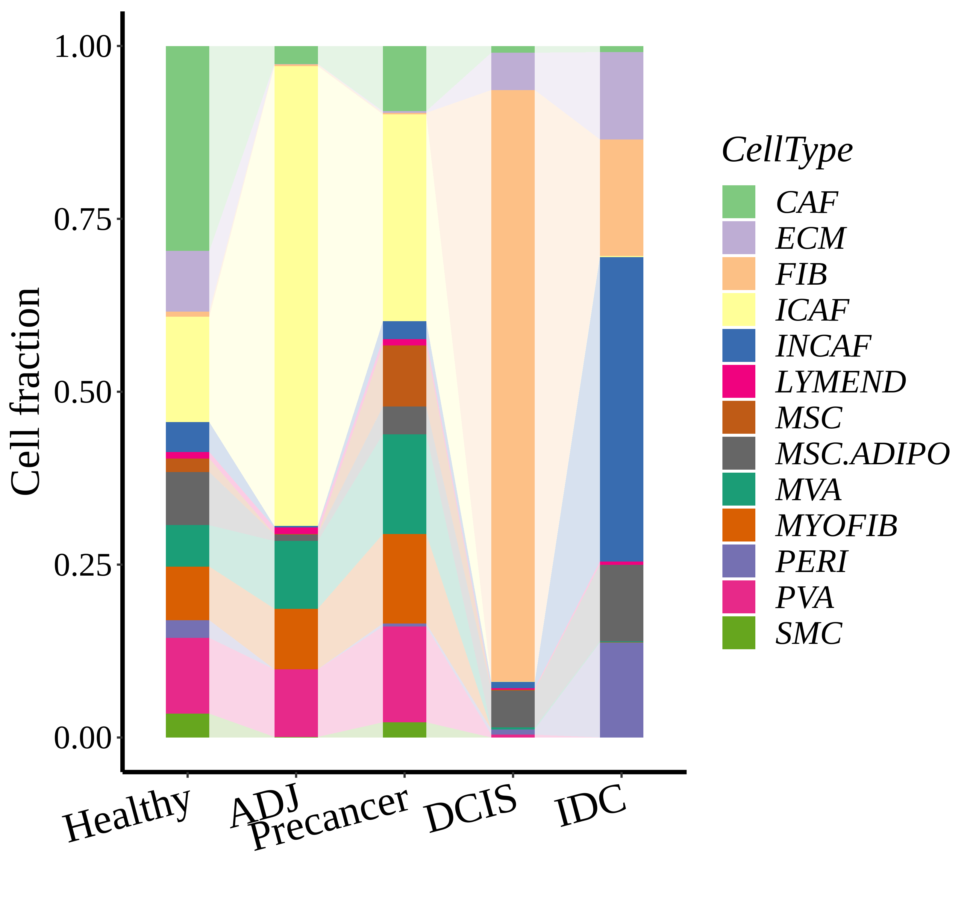
Stacked bar graph representation of the fraction of each epithelial cell type derived from samples with different Disease States in the integrated dataset of the selected tissue. Cells from each Disease State sample have a different color.Stacked bar graph representation of the fraction of each immune cell type derived from samples with different Disease States in the integrated dataset of the selected tissue. Cells from each Disease State sample have a different color.Stacked bar graph representation of the fraction of each stromal cell type derived from samples with different Disease States in the integrated dataset of the selected tissue. Cells from each Disease State sample have a different color.
∗Stacked bar graph representation of the fraction of each epithelial cell type derived from samples with different Disease States in the integrated dataset of Breast tissue. Cells from each Disease State sample have a different color.

Top

Cell type dynamics in malignant transformation

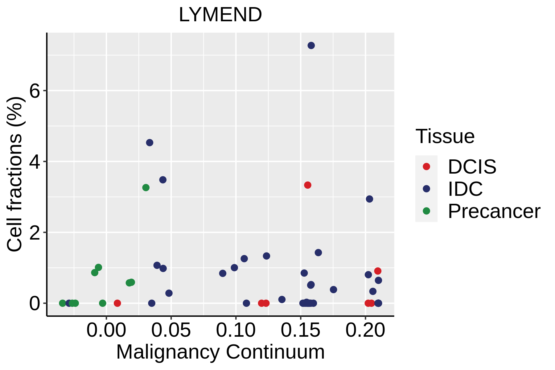
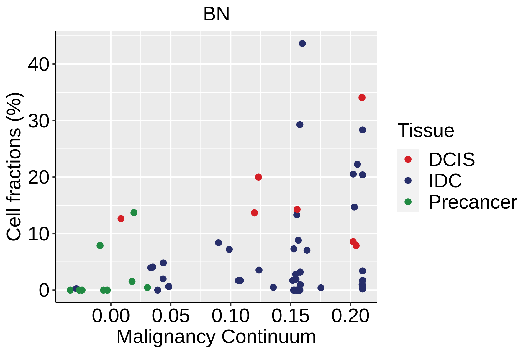
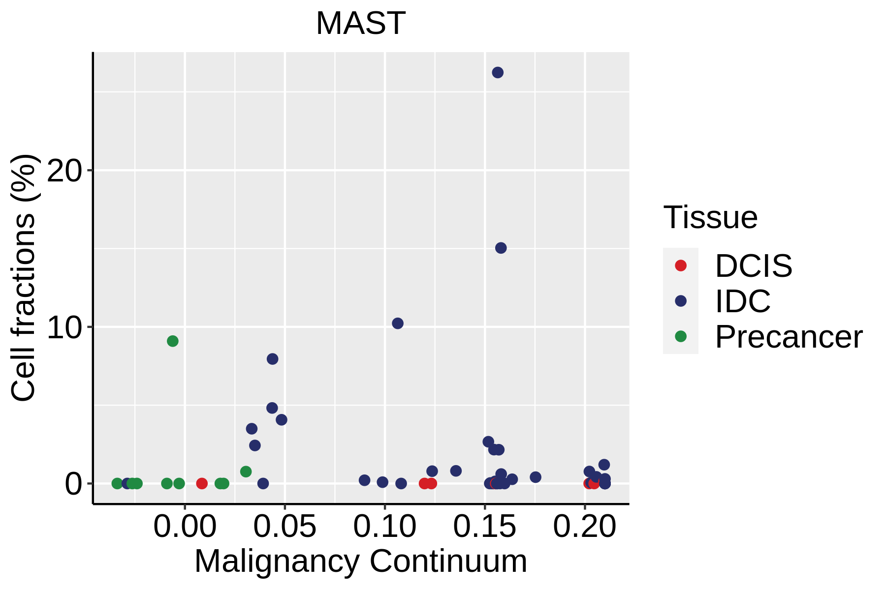
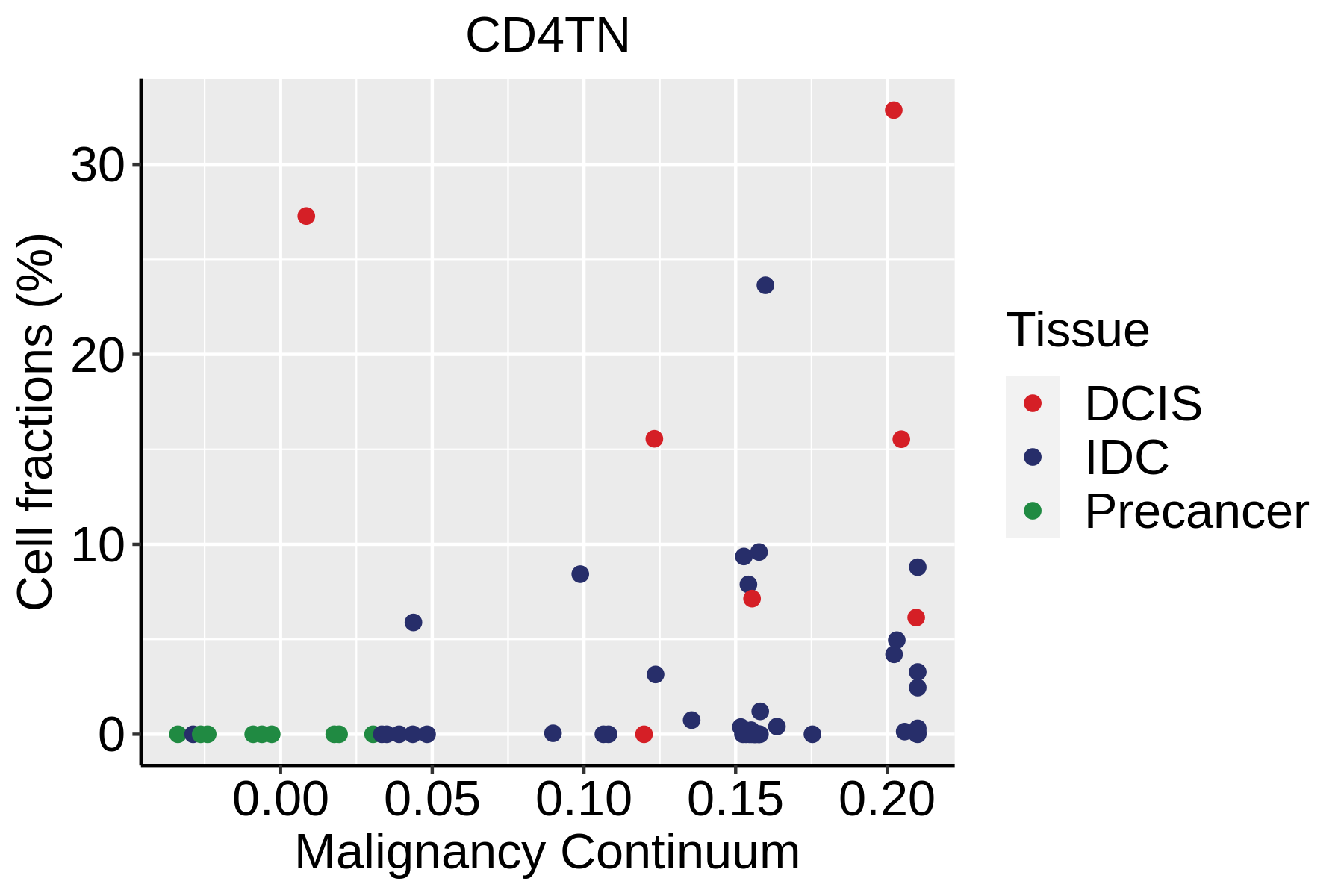
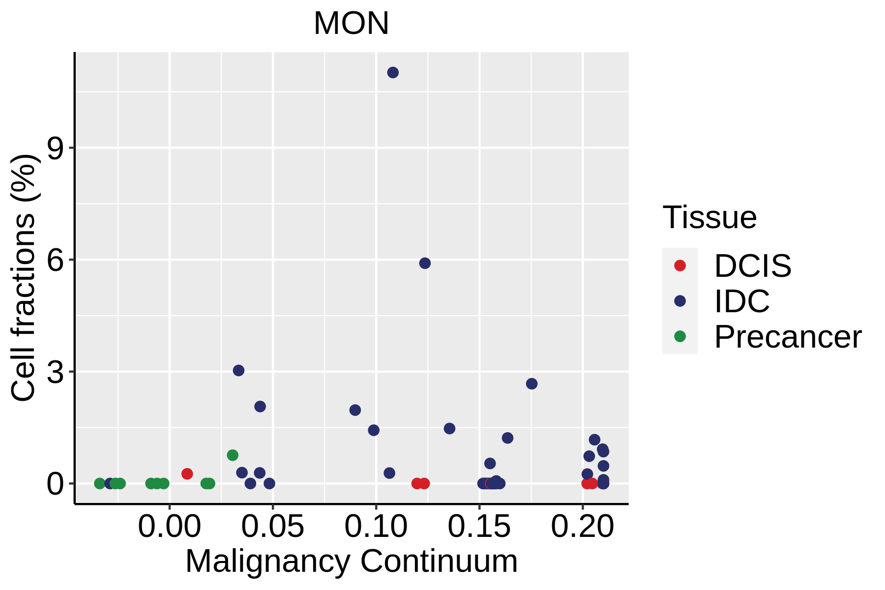
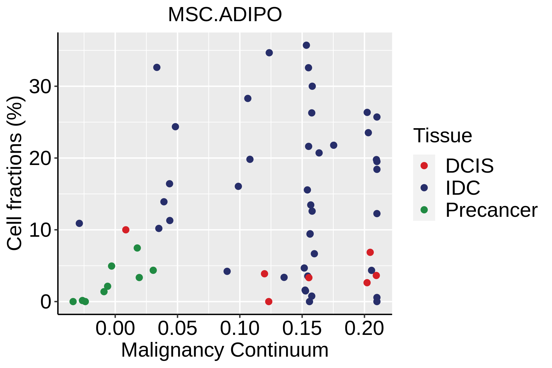
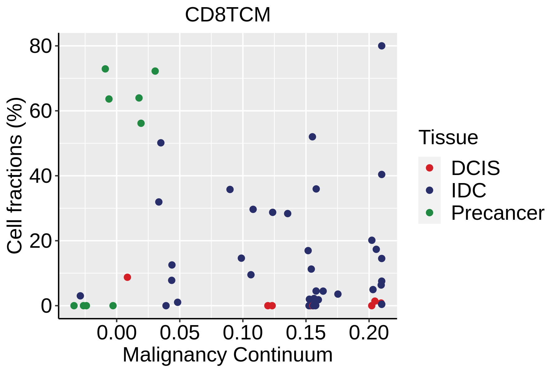
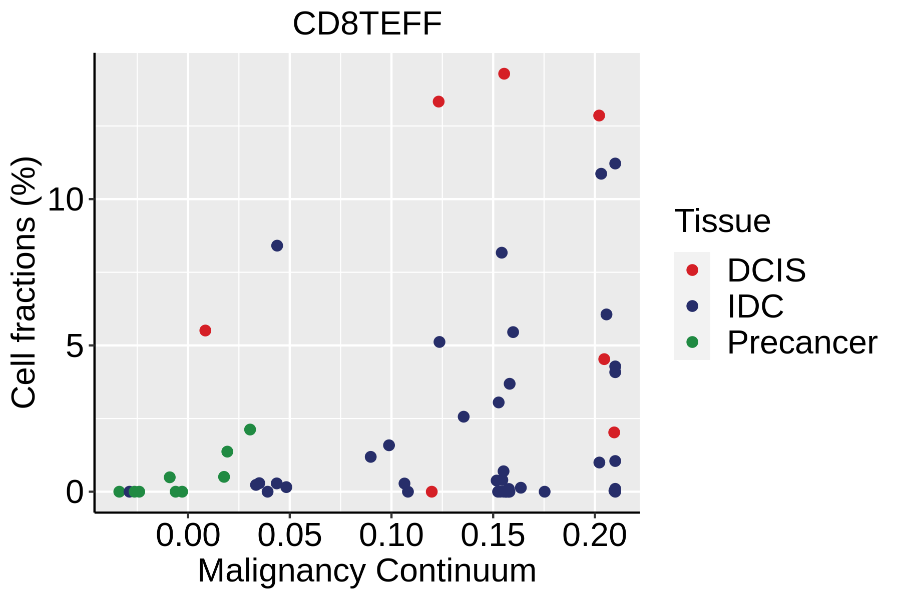
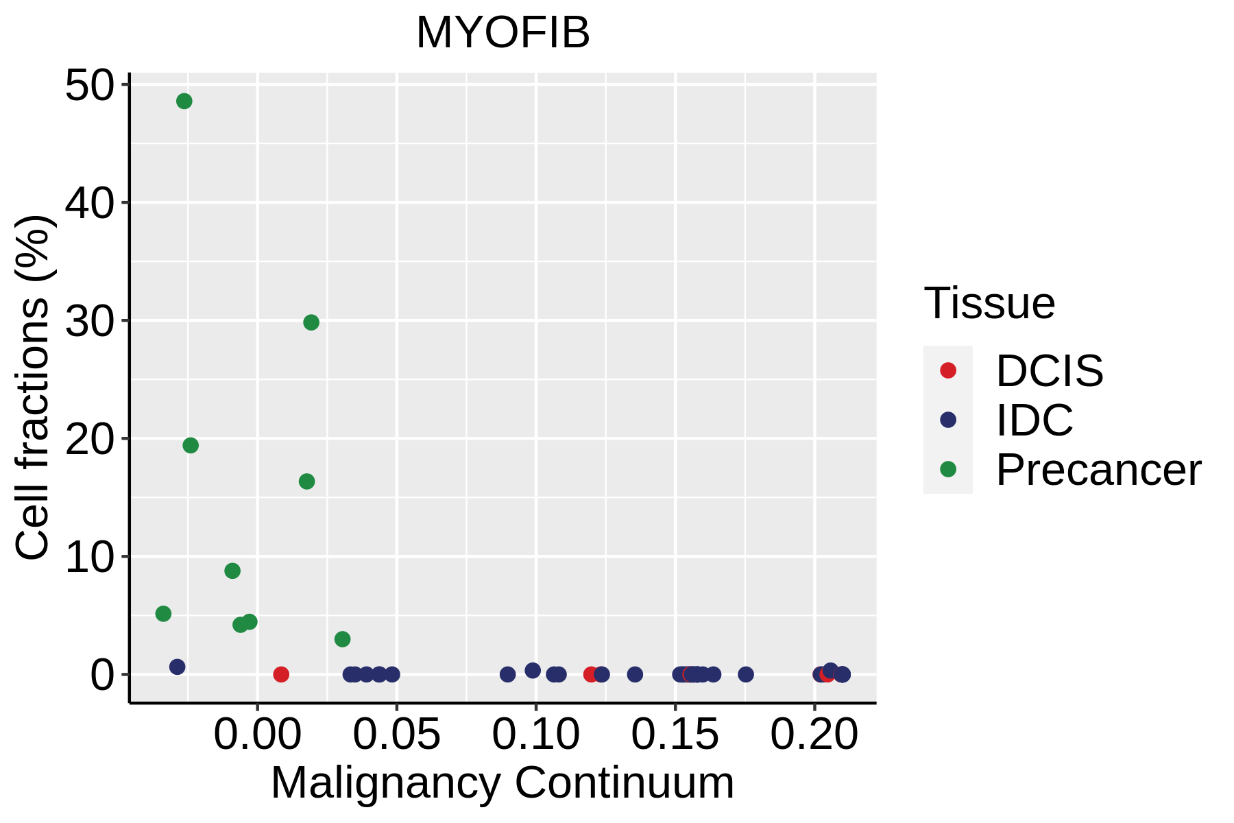
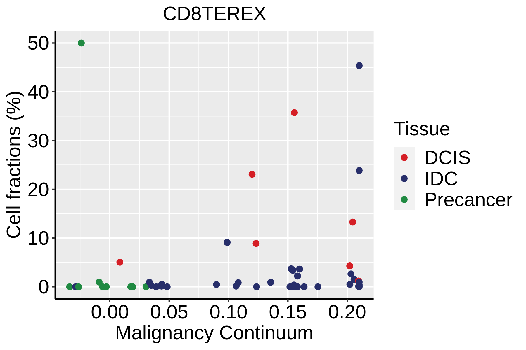
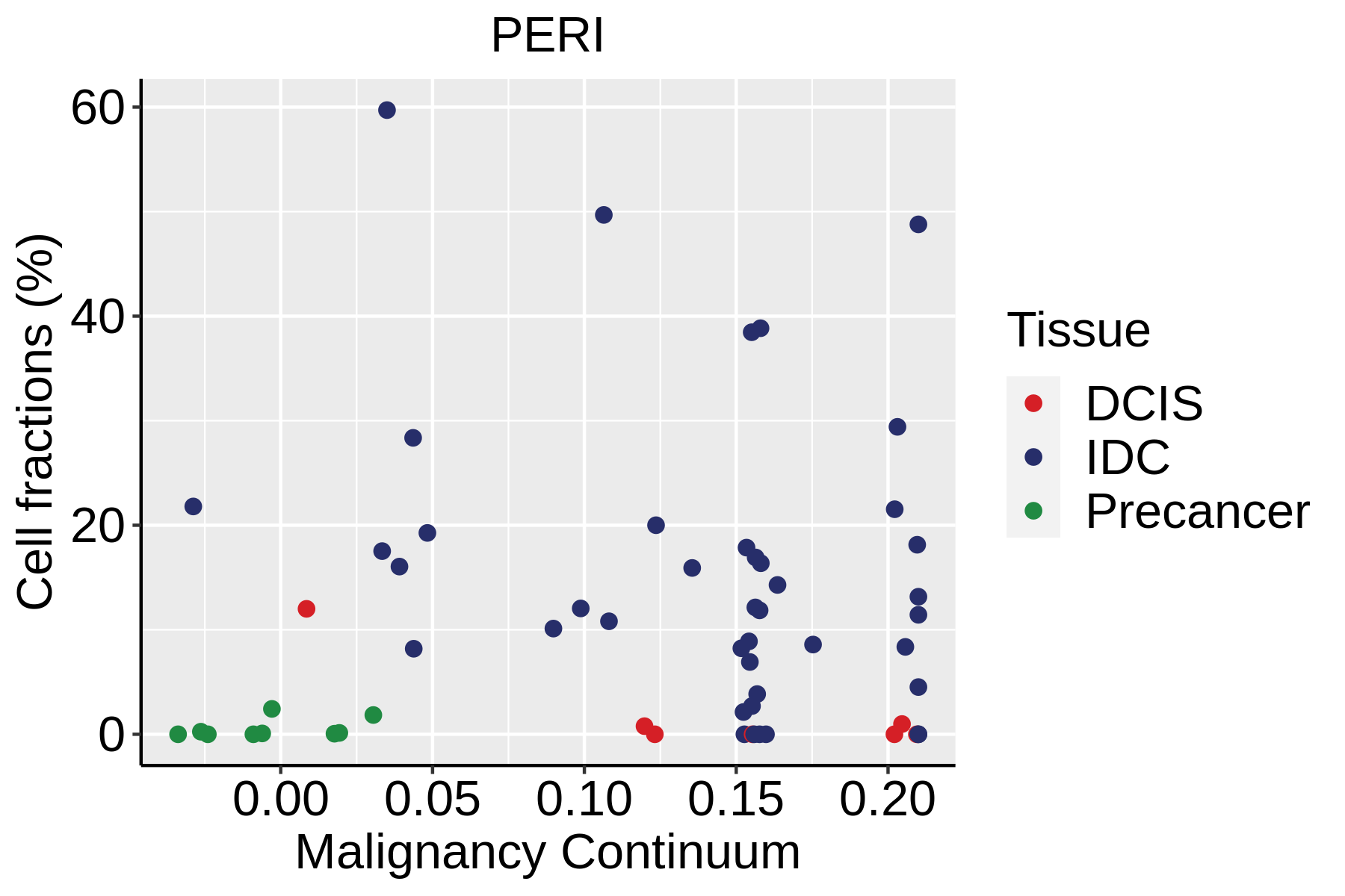
Fraction of cell type in each scRNA-seq sample plotted against position of the sample in the malignancy continuum for all cell types in the selected tissue. Samples are colored based on if they are derived from different Disease States.Fraction of cell type in each scRNA-seq sample plotted against position of the sample in the malignancy continuum for all cell types in the selected tissue. Samples are colored based on if they are derived from different Disease States.Fraction of cell type in each scRNA-seq sample plotted against position of the sample in the malignancy continuum for all cell types in the selected tissue. Samples are colored based on if they are derived from different Disease States.
∗Fraction of cell type in each scRNA-seq sample plotted against position of the sample in the malignancy continuum for all cell types in Breast tissue. Samples are colored based on if they are derived from different Disease States. Cell fractions are computed by dividing the number of cells of a given cell type by the total number of cells in the compartment (epithelial versus immune versus stromal).
Page: 1 2 3 

Top

Cell lineage trajectory inference

Disease StateEpitheliaImmuneStromal
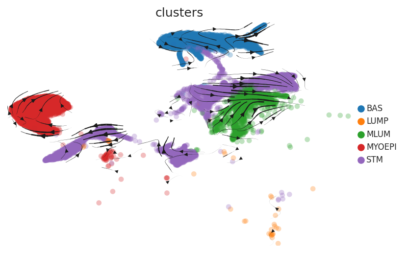
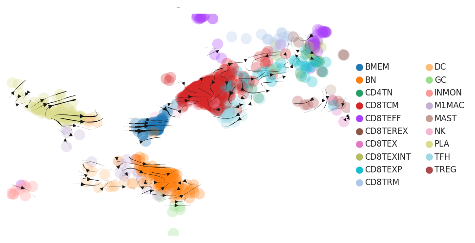
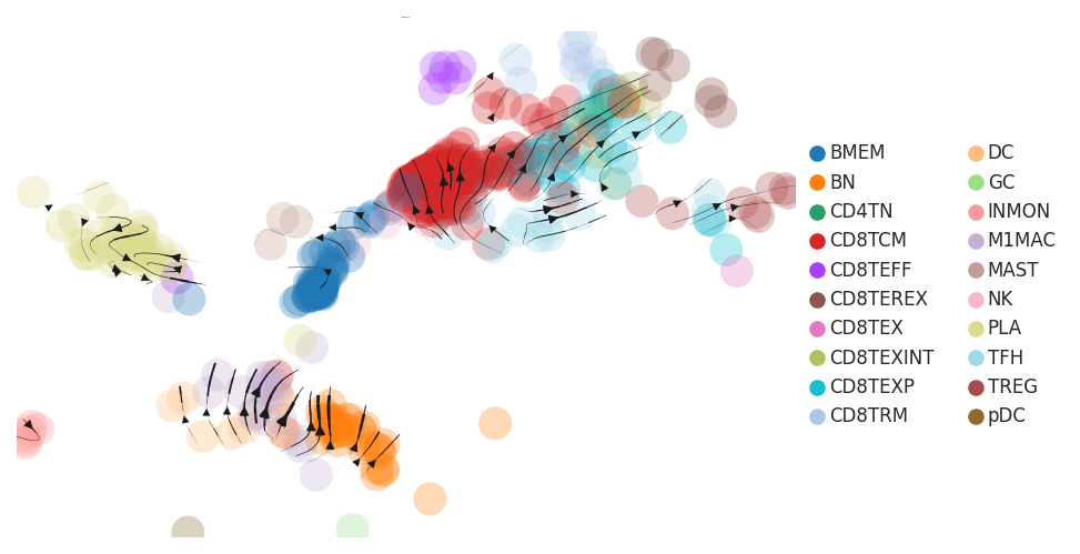
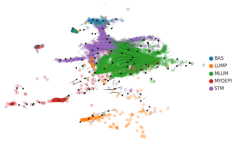
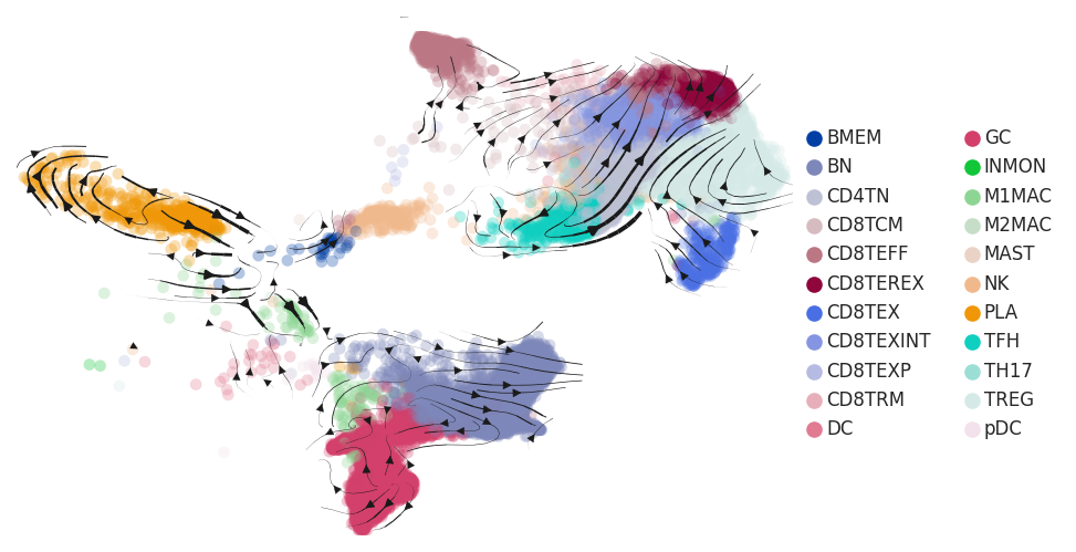
HealthyTrajectory analysis of cells of epithelial compartment in Healthy-derived samples of Breast tissue by using scvelo package in Python.Trajectory analysis of cells of immune compartment in Healthy-derived samples of Breast tissue by using scvelo package in Python.Trajectory analysis of cells of stromal compartment in Healthy-derived samples of Breast tissue by using scvelo package in Python.
ADJTrajectory analysis of cells of epithelial compartment in ADJ-derived samples of Breast tissue by using scvelo package in Python.Trajectory analysis of cells of immune compartment in ADJ-derived samples of Breast tissue by using scvelo package in Python.Trajectory analysis of cells of stromal compartment in ADJ-derived samples of Breast tissue by using scvelo package in Python.
PrecancerTrajectory analysis of cells of epithelial compartment in Precancer-derived samples of Breast tissue by using scvelo package in Python.Trajectory analysis of cells of immune compartment in Precancer-derived samples of Breast tissue by using scvelo package in Python.Trajectory analysis of cells of stromal compartment in Precancer-derived samples of Breast tissue by using scvelo package in Python.
DCISTrajectory analysis of cells of epithelial compartment in DCIS-derived samples of Breast tissue by using scvelo package in Python.Trajectory analysis of cells of immune compartment in DCIS-derived samples of Breast tissue by using scvelo package in Python.Trajectory analysis of cells of stromal compartment in DCIS-derived samples of Breast tissue by using scvelo package in Python.
IDCTrajectory analysis of cells of epithelial compartment in IDC-derived samples of Breast tissue by using scvelo package in Python.Trajectory analysis of cells of immune compartment in IDC-derived samples of Breast tissue by using scvelo package in Python.Trajectory analysis of cells of stromal compartment in IDC-derived samples of Breast tissue by using scvelo package in Python.
∗RNA velocity maps for epithelial, immune and stromal compartments of Breast tissue with different disease states.

Top

TF regulatory network analysis

Disease StateEpitheliaImmuneStromal
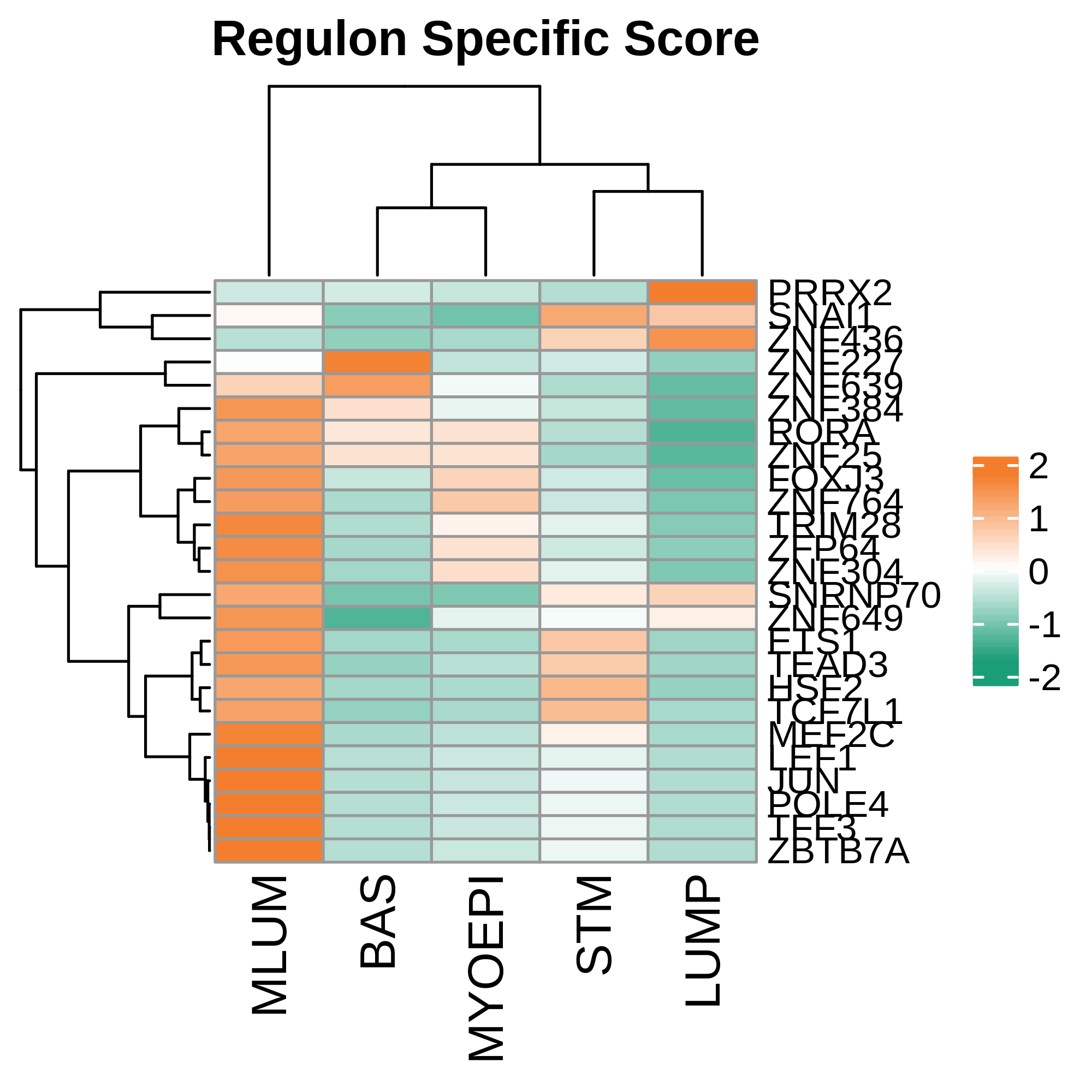
HealthyHeatmap representation of significantly different TFs among all cell types of epithelial compartment in Healthy-derived samples of Breast tissue by comparing their average AUC by using pySCENIC in Python.Heatmap representation of significantly different TFs among all cell types of immune compartment in Healthy-derived samples of Breast tissue by comparing their average AUC by using pySCENIC in Python.Heatmap representation of significantly different TFs among all cell types of stromal compartment in Healthy-derived samples of Breast tissue by comparing their average AUC by using pySCENIC in Python.
TF target networkTF target networkTF target network
ADJHeatmap representation of significantly different TFs among all cell types of epithelial compartment in ADJ-derived samples of Breast tissue by comparing their average AUC by using pySCENIC in Python.Heatmap representation of significantly different TFs among all cell types of immune compartment in ADJ-derived samples of Breast tissue by comparing their average AUC by using pySCENIC in Python.Heatmap representation of significantly different TFs among all cell types of stromal compartment in ADJ-derived samples of Breast tissue by comparing their average AUC by using pySCENIC in Python.
TF target networkTF target networkTF target network
PrecancerHeatmap representation of significantly different TFs among all cell types of epithelial compartment in Precancer-derived samples of Breast tissue by comparing their average AUC by using pySCENIC in Python.Heatmap representation of significantly different TFs among all cell types of immune compartment in Precancer-derived samples of Breast tissue by comparing their average AUC by using pySCENIC in Python.Heatmap representation of significantly different TFs among all cell types of stromal compartment in Precancer-derived samples of Breast tissue by comparing their average AUC by using pySCENIC in Python.
TF target networkTF target networkTF target network
DCISHeatmap representation of significantly different TFs among all cell types of epithelial compartment in DCIS-derived samples of Breast tissue by comparing their average AUC by using pySCENIC in Python.Heatmap representation of significantly different TFs among all cell types of immune compartment in DCIS-derived samples of Breast tissue by comparing their average AUC by using pySCENIC in Python.Heatmap representation of significantly different TFs among all cell types of stromal compartment in DCIS-derived samples of Breast tissue by comparing their average AUC by using pySCENIC in Python.
TF target networkTF target networkTF target network
IDCHeatmap representation of significantly different TFs among all cell types of epithelial compartment in IDC-derived samples of Breast tissue by comparing their average AUC by using pySCENIC in Python.Heatmap representation of significantly different TFs among all cell types of immune compartment in IDC-derived samples of Breast tissue by comparing their average AUC by using pySCENIC in Python.Heatmap representation of significantly different TFs among all cell types of stromal compartment in IDC-derived samples of Breast tissue by comparing their average AUC by using pySCENIC in Python.
TF target networkTF target networkTF target network
∗Heatmap: Heatmap representation of significantly different TFs among all cell types of epithelial compartment in Healthy-derived samples of Breast tissue by comparing their average AUC by using pySCENIC in Python. Regulon specific score for each cell type is colored.
∗Network: TF target network created from Healthy-derived samples of Breast tissue using Top 50 predicted target genes of each regulon. Edge lengths are the inferred TF-target weightings.

Top

Cell-cell interaction analysis

Disease StateCell TypeCircleSourceTarget
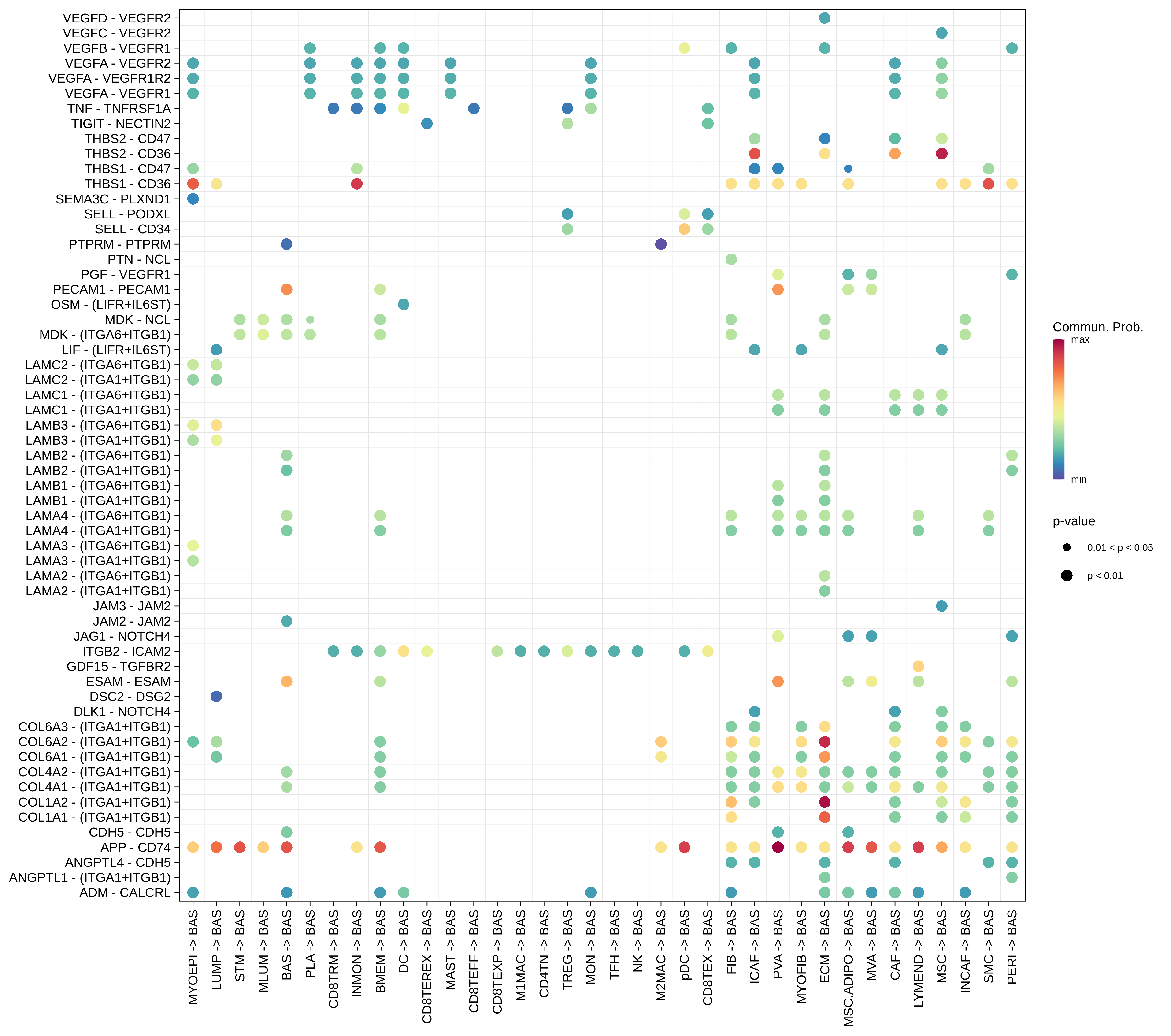
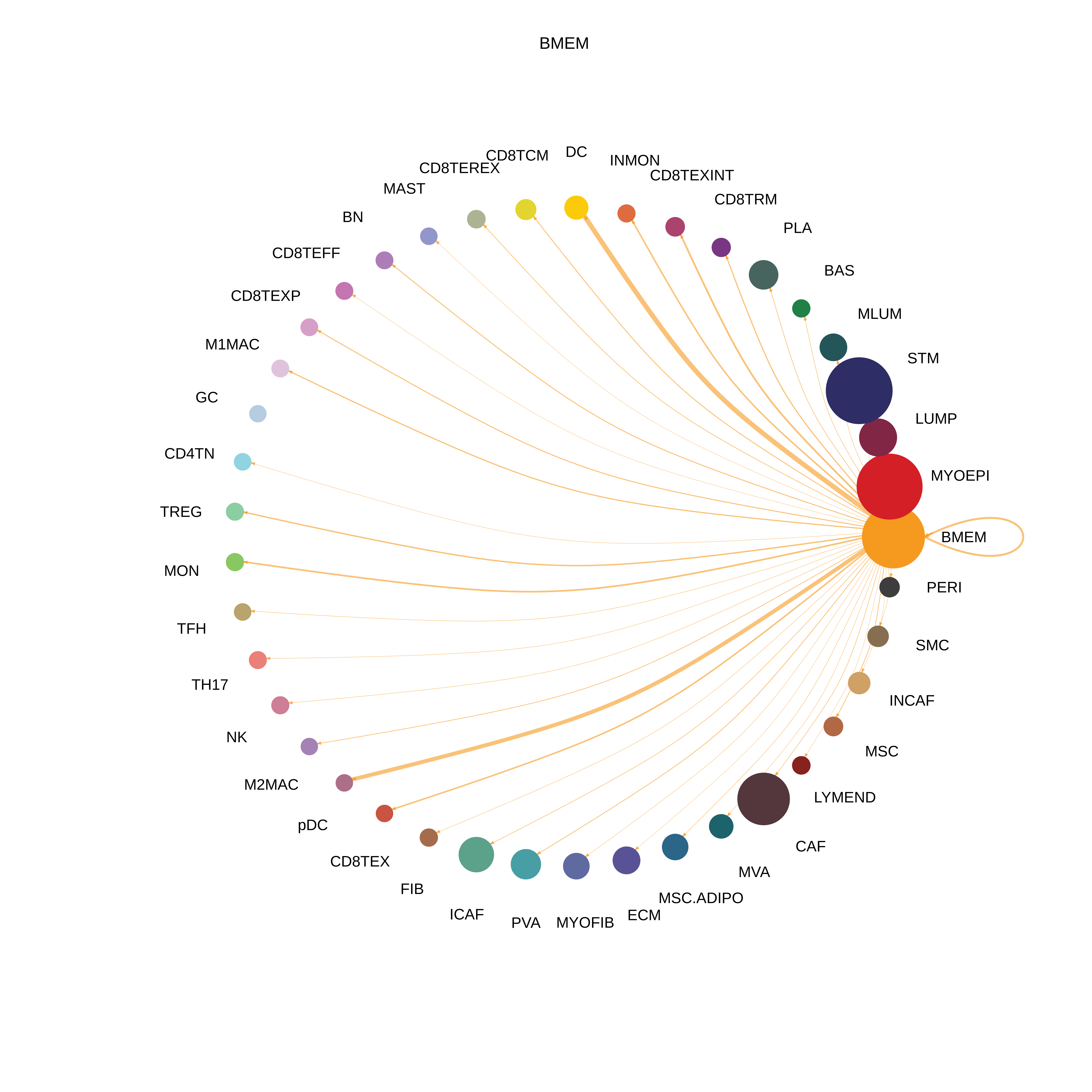
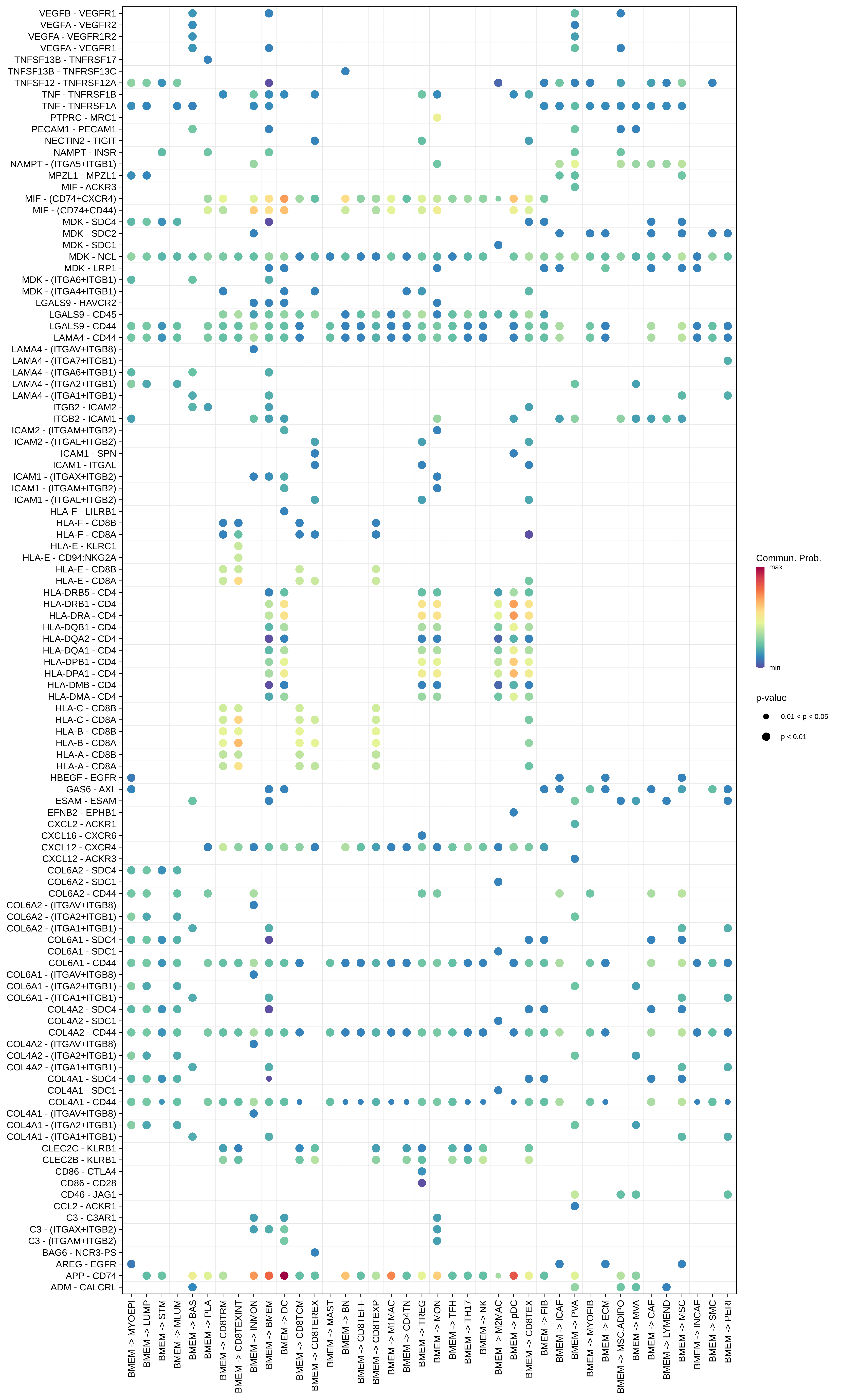
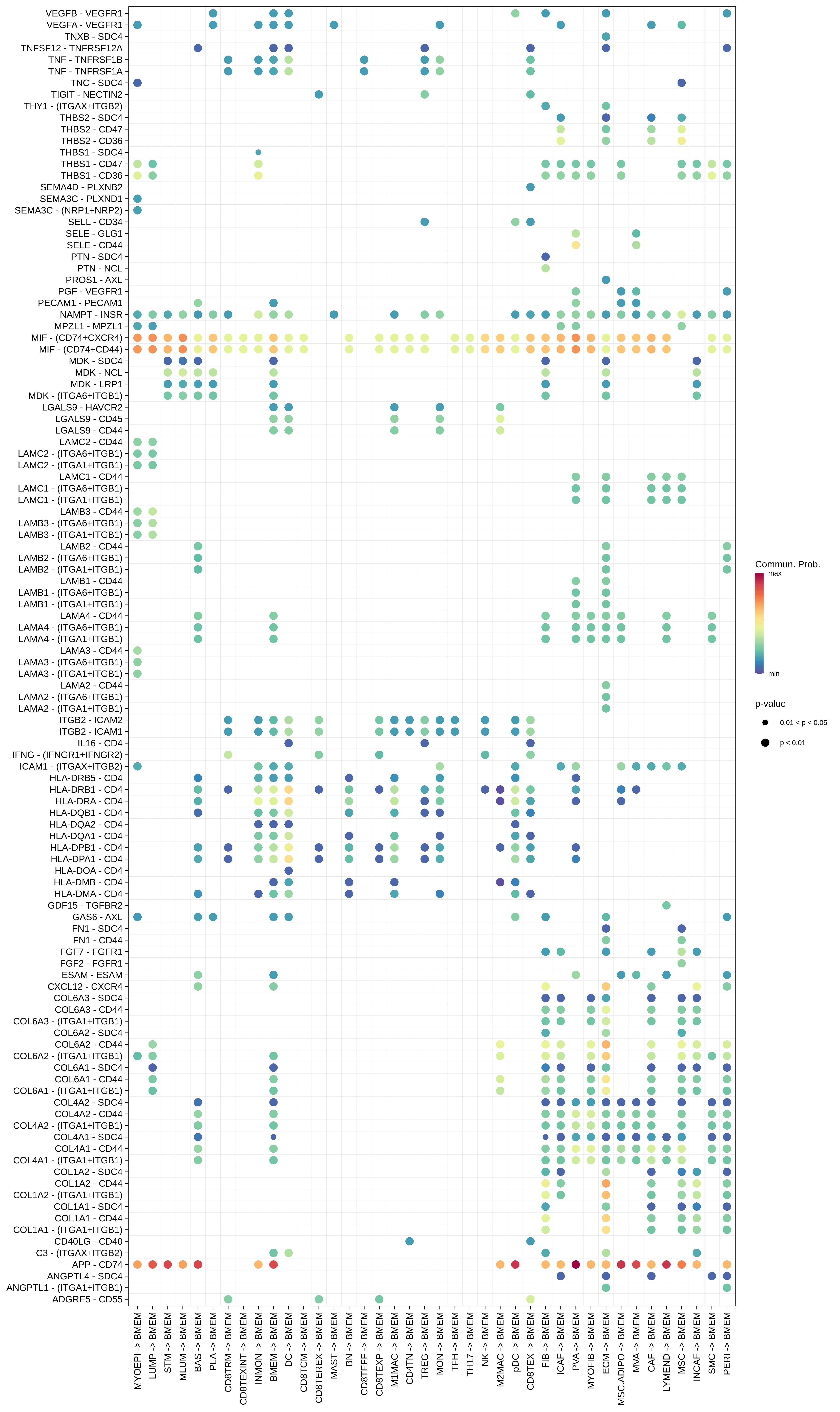
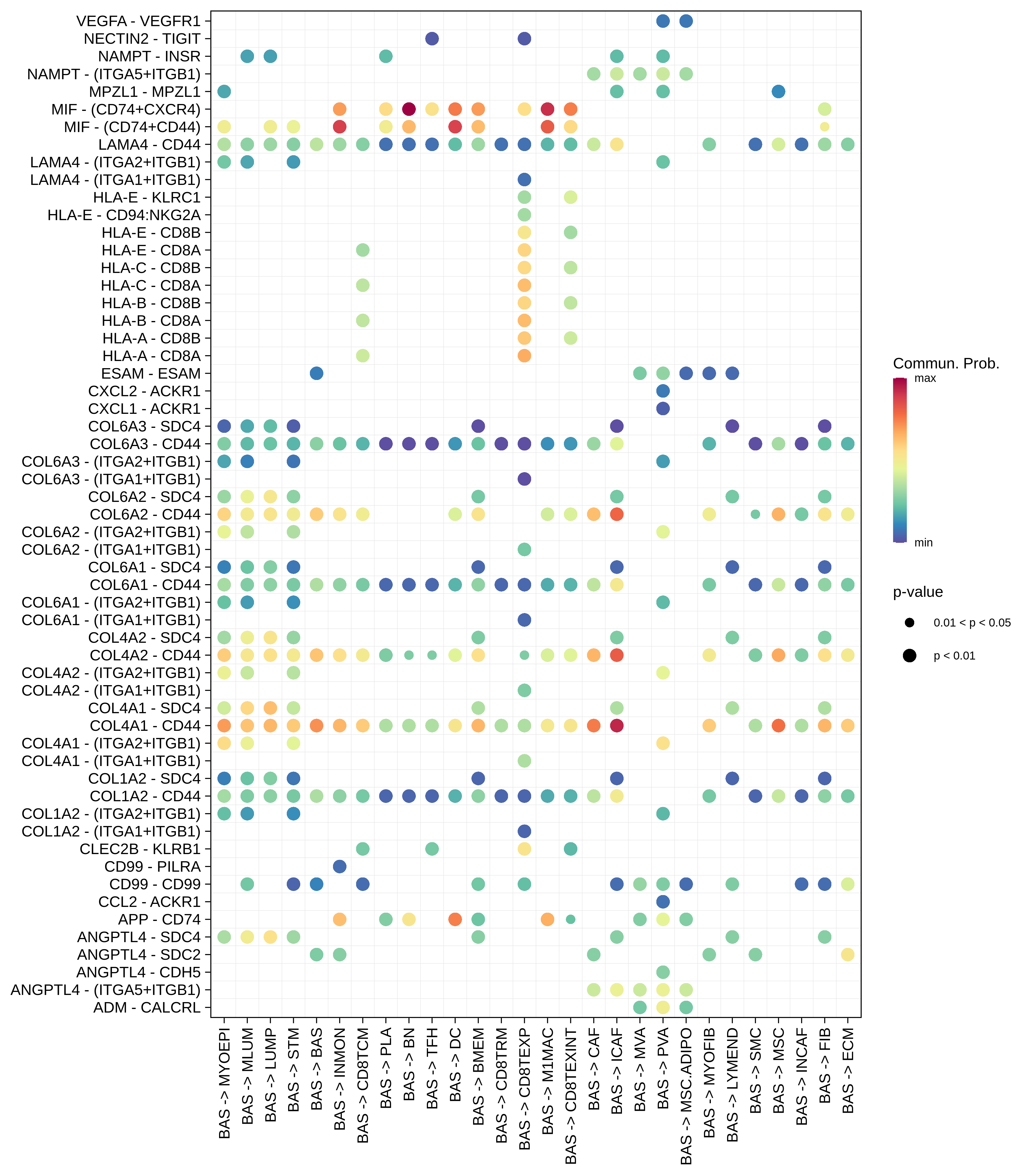
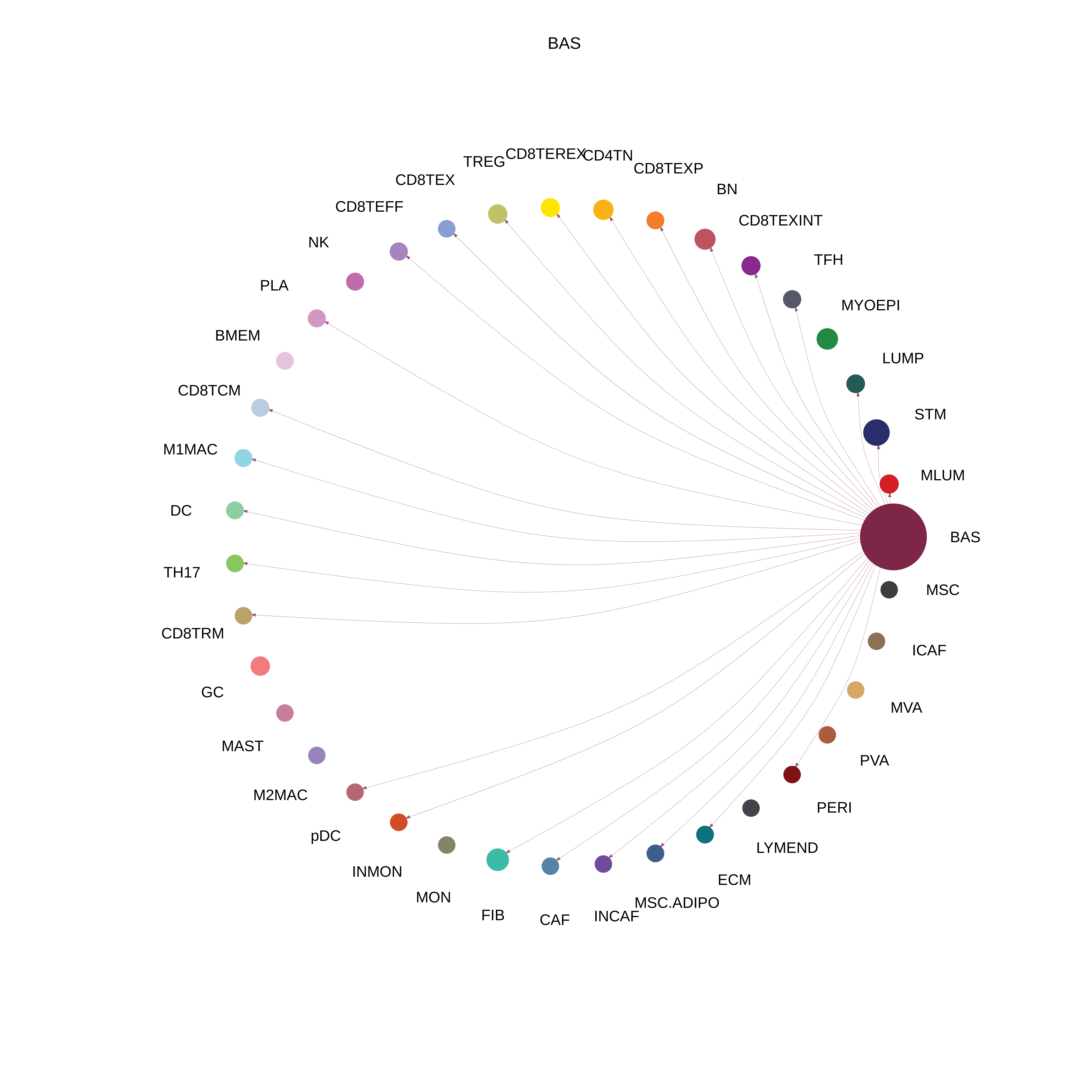
HealthyBASThe cell-cell communications between each two interacting cell types in Healthy-derived samples of the selected tissue. Different colors in the circle plot represent different cell groups and the edge width is proportional to the indicated number of ligand-receptor pairs.Bubble plot of proportion to the communication probability in significant signaling pathways between this cell type and its source cell population in Healthy-derived samples of the selected tissue. Each row represents the significant ligand and receptor gene pairs.Bubble plot of proportion to the communication probability in significant signaling pathways between this cell type and its target cell population in Healthy-derived samples of the selected tissue. Each row represents the significant ligand and receptor gene pairs.
BMEMThe cell-cell communications between each two interacting cell types in Healthy-derived samples of the selected tissue. Different colors in the circle plot represent different cell groups and the edge width is proportional to the indicated number of ligand-receptor pairs.Bubble plot of proportion to the communication probability in significant signaling pathways between this cell type and its source cell population in Healthy-derived samples of the selected tissue. Each row represents the significant ligand and receptor gene pairs.Bubble plot of proportion to the communication probability in significant signaling pathways between this cell type and its target cell population in Healthy-derived samples of the selected tissue. Each row represents the significant ligand and receptor gene pairs.
Page: 1 2 3 4 5 6 7 8 9 10 11 12 13 14 15 16 17 18 19 20 

Disease StateCell TypeCircleSourceTarget
ADJBASThe cell-cell communications between each two interacting cell types in ADJ-derived samples of the selected tissue. Different colors in the circle plot represent different cell groups and the edge width is proportional to the indicated number of ligand-receptor pairs. Bubble plot of proportion to the communication probability in significant signaling pathways between this cell type and its source cell population in ADJ-derived samples of the selected tissue. Each row represents the significant ligand and receptor gene pairs.Bubble plot of proportion to the communication probability in significant signaling pathways between this cell type and its target cell population in ADJ-derived samples of the selected tissue. Each row represents the significant ligand and receptor gene pairs.
BMEMThe cell-cell communications between each two interacting cell types in ADJ-derived samples of the selected tissue. Different colors in the circle plot represent different cell groups and the edge width is proportional to the indicated number of ligand-receptor pairs. Bubble plot of proportion to the communication probability in significant signaling pathways between this cell type and its source cell population in ADJ-derived samples of the selected tissue. Each row represents the significant ligand and receptor gene pairs.Bubble plot of proportion to the communication probability in significant signaling pathways between this cell type and its target cell population in ADJ-derived samples of the selected tissue. Each row represents the significant ligand and receptor gene pairs.
Page: 1 2 3 4 5 6 7 8 9 10 11 12 13 14 15 

Disease StateCell TypeCircleSourceTarget
PrecancerBASThe cell-cell communications between each two interacting cell types in Precancer-derived samples of the selected tissue. Different colors in the circle plot represent different cell groups and the edge width is proportional to the indicated number of ligand-receptor pairs. Bubble plot of proportion to the communication probability in significant signaling pathways between this cell type and its source cell population in Precancer-derived samples of the selected tissue. Each row represents the significant ligand and receptor gene pairs.Bubble plot of proportion to the communication probability in significant signaling pathways between this cell type and its target cell population in Precancer-derived samples of the selected tissue. Each row represents the significant ligand and receptor gene pairs.
BMEMThe cell-cell communications between each two interacting cell types in Precancer-derived samples of the selected tissue. Different colors in the circle plot represent different cell groups and the edge width is proportional to the indicated number of ligand-receptor pairs. Bubble plot of proportion to the communication probability in significant signaling pathways between this cell type and its source cell population in Precancer-derived samples of the selected tissue. Each row represents the significant ligand and receptor gene pairs.Bubble plot of proportion to the communication probability in significant signaling pathways between this cell type and its target cell population in Precancer-derived samples of the selected tissue. Each row represents the significant ligand and receptor gene pairs.
Page: 1 2 3 4 5 6 7 8 9 10 11 12 13 14 15 

Disease StateCell TypeCircleSourceTarget
DCISBASThe cell-cell communications between each two interacting cell types in DCIS-derived samples of Breast tissue. Different colors in the circle plot represent different cell groups and the edge width is proportional to the indicated number of ligand-receptor pairs. Bubble plot of proportion to the communication probability in significant signaling pathways between this cell type and its source cell population in DCIS-derived samples of Breast tissue. Each row represents the significant ligand and receptor gene pairs.Bubble plot of proportion to the communication probability in significant signaling pathways between this cell type and its target cell population in DCIS-derived samples of Breast tissue. Each row represents the significant ligand and receptor gene pairs.
BMEMThe cell-cell communications between each two interacting cell types in DCIS-derived samples of Breast tissue. Different colors in the circle plot represent different cell groups and the edge width is proportional to the indicated number of ligand-receptor pairs. Bubble plot of proportion to the communication probability in significant signaling pathways between this cell type and its source cell population in DCIS-derived samples of Breast tissue. Each row represents the significant ligand and receptor gene pairs.Bubble plot of proportion to the communication probability in significant signaling pathways between this cell type and its target cell population in DCIS-derived samples of Breast tissue. Each row represents the significant ligand and receptor gene pairs.
Page: 1 2 3 4 5 6 7 8 9 10 11 12 13 14 15 16 

Disease StateCell TypeCircleSourceTarget
IDCBASThe cell-cell communications between each two interacting cell types in IDC-derived samples of Breast tissue. Different colors in the circle plot represent different cell groups and the edge width is proportional to the indicated number of ligand-receptor pairs. Bubble plot of proportion to the communication probability in significant signaling pathways between this cell type and its source cell population in IDC-derived samples of Breast tissue. Each row represents the significant ligand and receptor gene pairs.Bubble plot of proportion to the communication probability in significant signaling pathways between this cell type and its target cell population in IDC-derived samples of Breast tissue. Each row represents the significant ligand and receptor gene pairs.
BMEMThe cell-cell communications between each two interacting cell types in IDC-derived samples of Breast tissue. Different colors in the circle plot represent different cell groups and the edge width is proportional to the indicated number of ligand-receptor pairs. Bubble plot of proportion to the communication probability in significant signaling pathways between this cell type and its source cell population in IDC-derived samples of Breast tissue. Each row represents the significant ligand and receptor gene pairs.Bubble plot of proportion to the communication probability in significant signaling pathways between this cell type and its target cell population in IDC-derived samples of Breast tissue. Each row represents the significant ligand and receptor gene pairs.
∗The cell-cell communications between each two interacting cell types in different states-derived samples of Breast tissue. Different colors in the circle plot represent different cell groups and the edge width is proportional to the indicated number of ligand-receptor pairs.
∗Bubble plot of proportion to the communication probability in significant signaling pathways between this cell type and its source and target cell population. Each row represents the significant ligand and receptor gene pairs.
Page: 1 2 3 4 5 6 7 8 9 10 11 12 13 14 15 16 17 18 19 

Top

Stemness, Senescence, Metaplasia and NeoTCR signature analysis

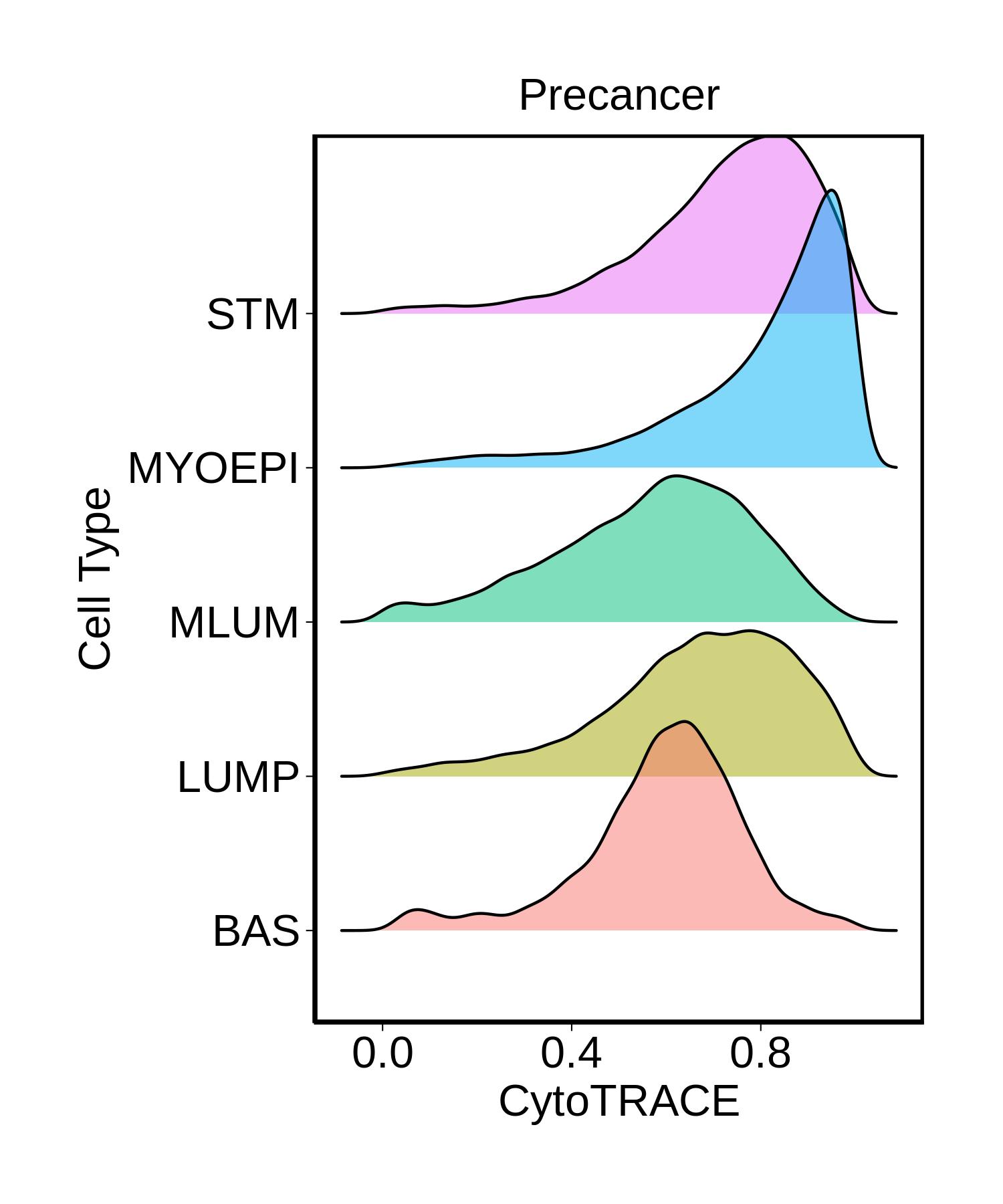
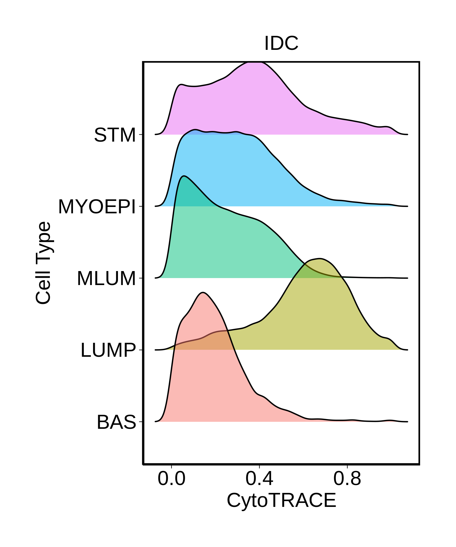
check button Developmental potential inference using CytoTRACE.
1_developmental_potential_in_Healthy.png shows the analysis results of stemness, senescence, and metaplasia signatures2_developmental_potential_in_Precancer.png shows the analysis results of stemness, senescence, and metaplasia signatures3_developmental_potential_in_DCIS.png shows the analysis results of stemness, senescence, and metaplasia signatures4_developmental_potential_in_IDC.png shows the analysis results of stemness, senescence, and metaplasia signatures
∗Ridge plots of CytoTRACE score distributions for epithelial cells.

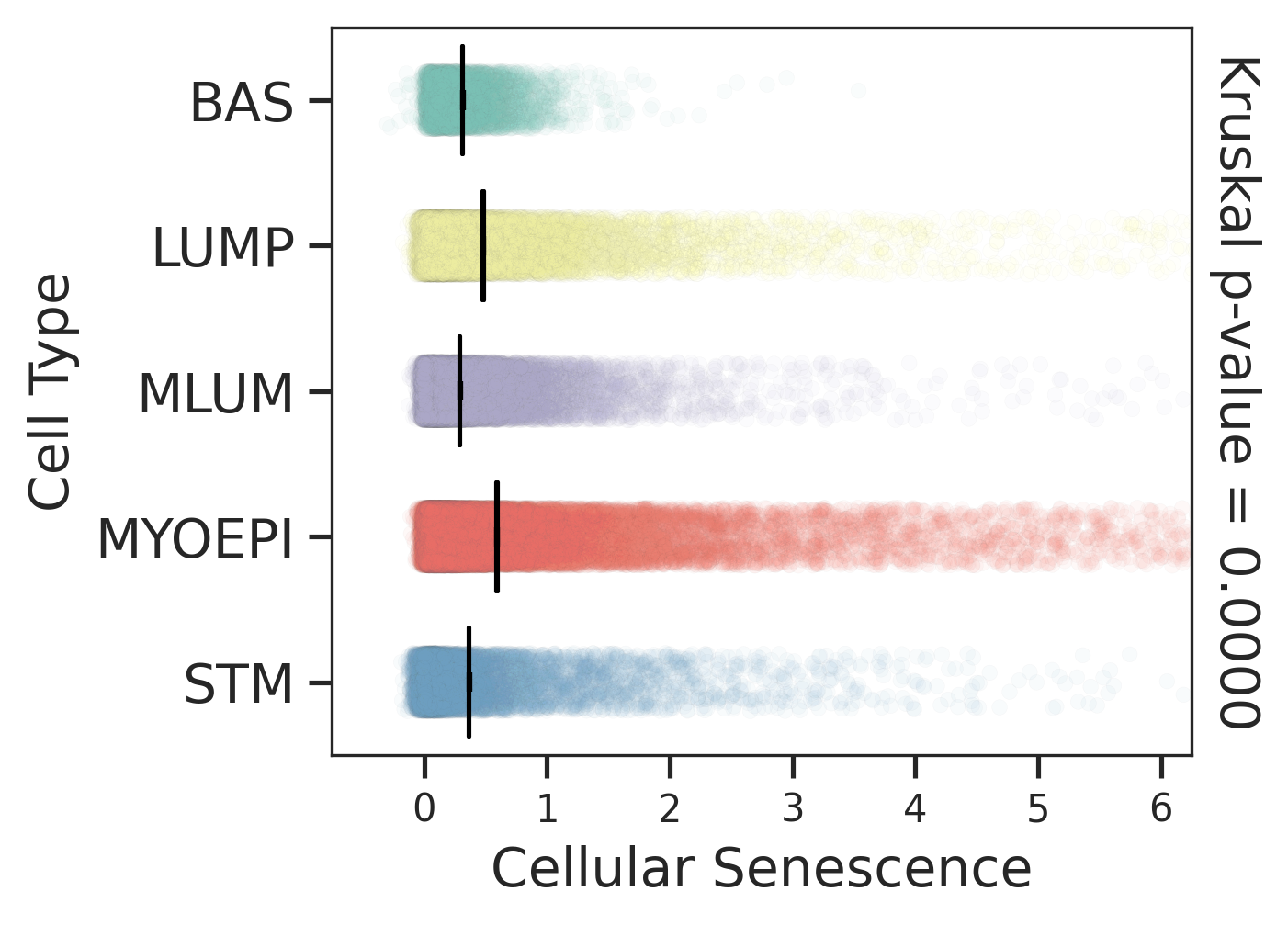
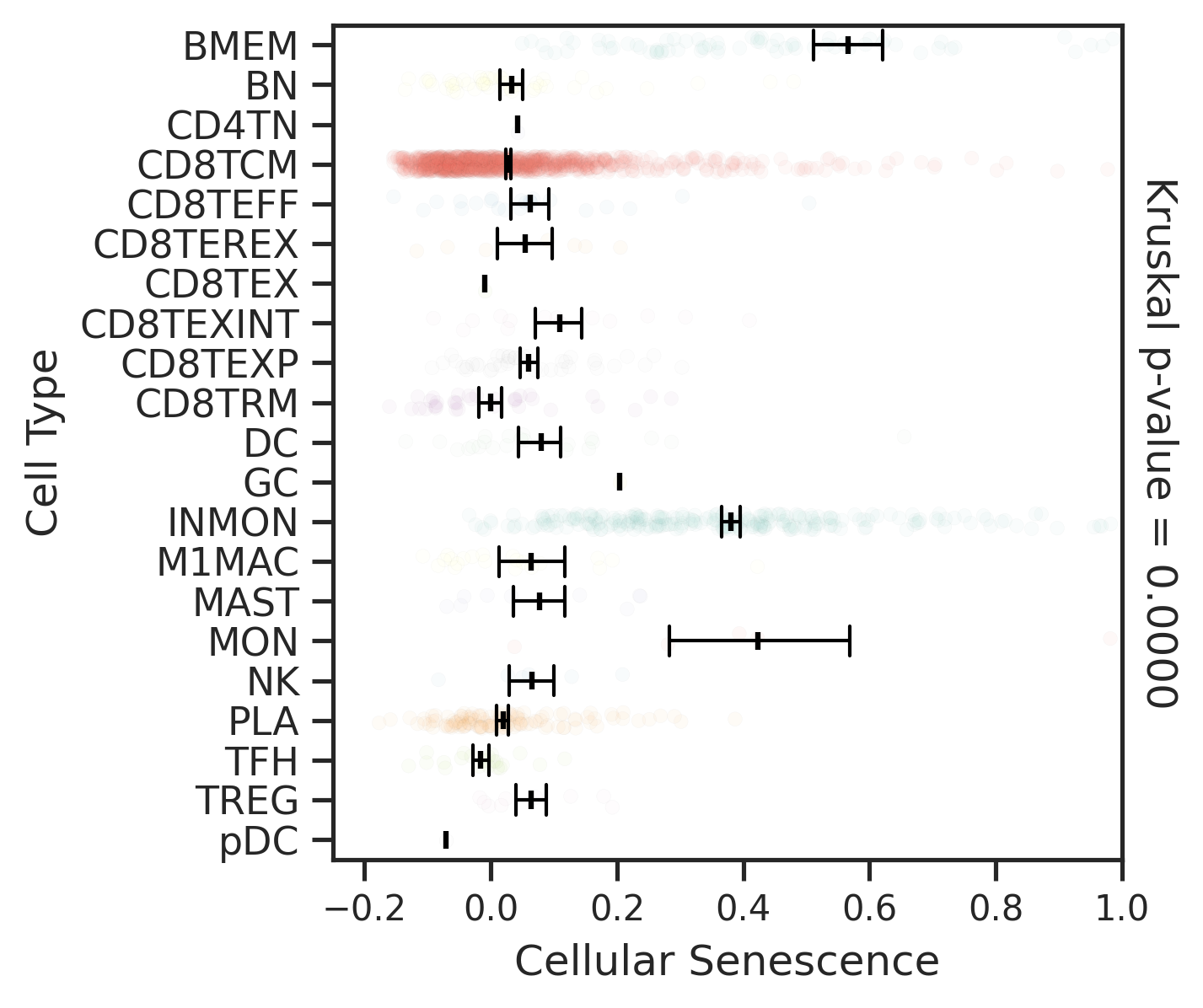
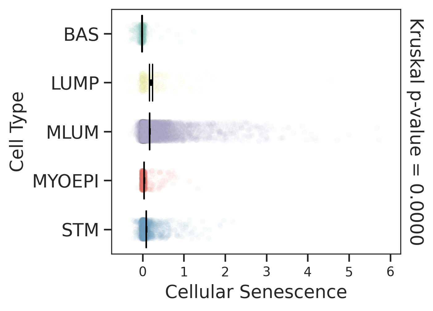
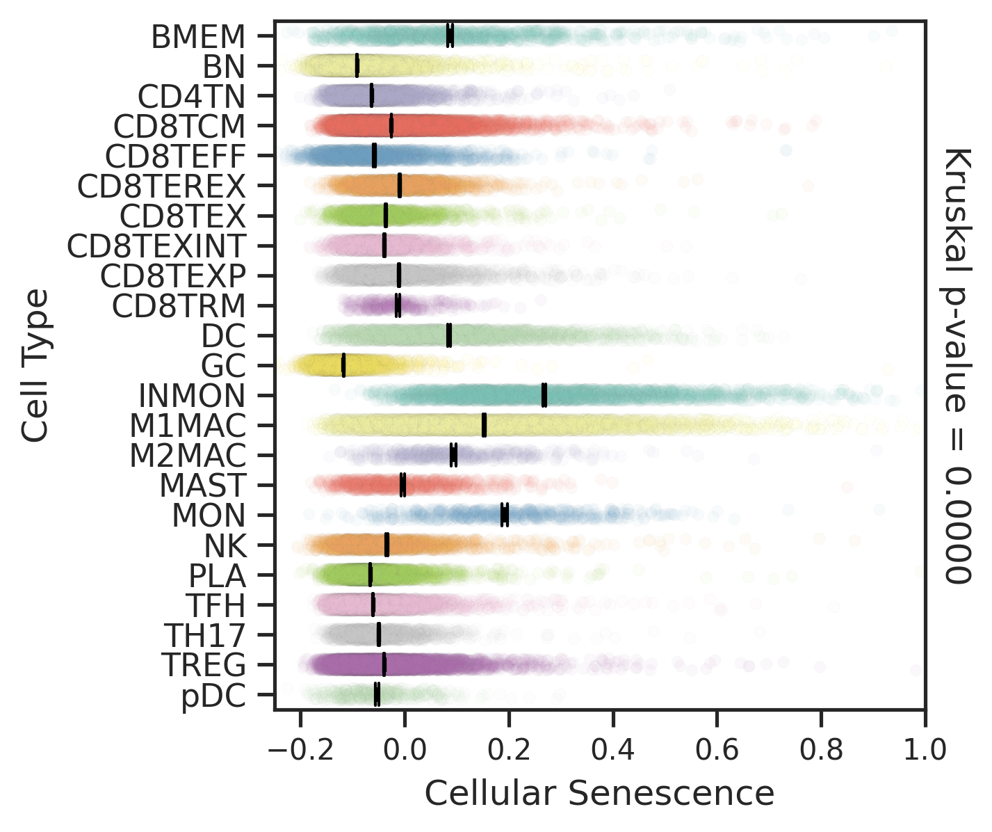
check button Cellular senescence signature (for three compartments).
Disease StateEpitheliaImmuneStromal
HealthyComparisons for degree of cellular senescence of epithelial compartment in Healthy-derived samples of Breast tissue.Comparisons for degree of cellular senescence of immune compartment in Healthy-derived samples of Breast tissue.Comparisons for degree of cellular senescence of stromal compartment in Healthy-derived samples of Breast tissue.
PrecancerComparisons for degree of cellular senescence of epithelial compartment in Precancer-derived samples of Breast tissue.Comparisons for degree of cellular senescence of immune compartment in Precancer-derived samples of Breast tissue.Comparisons for degree of cellular senescence of stromal compartment in Precancer-derived samples of Breast tissue.
DCISComparisons for degree of cellular senescence of epithelial compartment in DCIS-derived samples of Breast tissue.Comparisons for degree of cellular senescence of immune compartment in DCIS-derived samples of Breast tissue.Comparisons for degree of cellular senescence of stromal compartment in DCIS-derived samples of Breast tissue.
IDCComparisons for degree of cellular senescence of epithelial compartment in IDC-derived samples of Breast tissue.Comparisons for degree of cellular senescence of immune compartment in IDC-derived samples of Breast tissue.Comparisons for degree of cellular senescence of stromal compartment in IDC-derived samples of Breast tissue.
∗Comparisons for degree of cellular senescence among epithelial, immune and stromal cell populations of Breast tissue with different disease states.

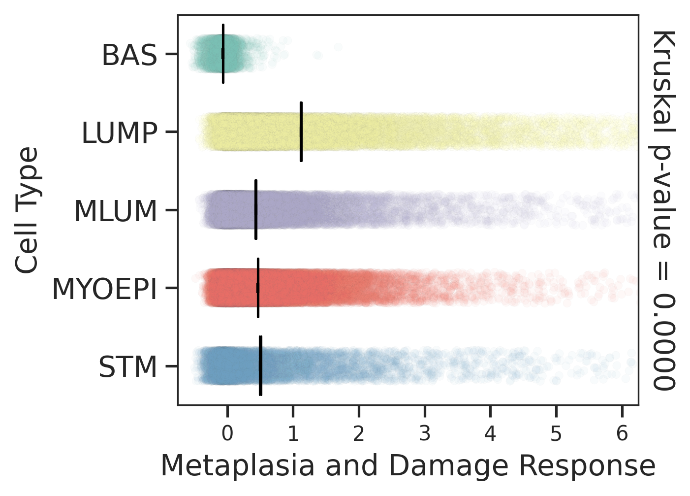
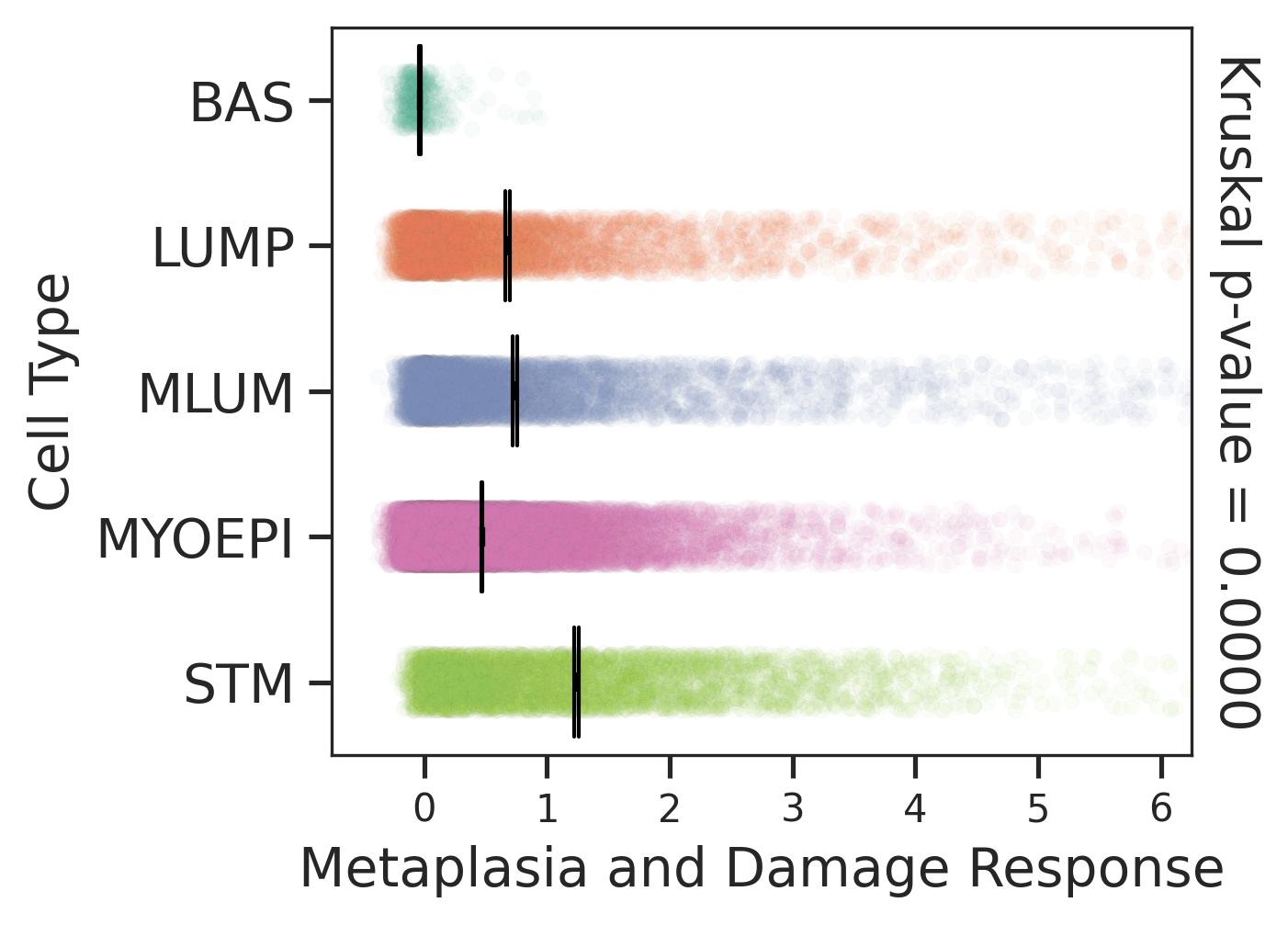
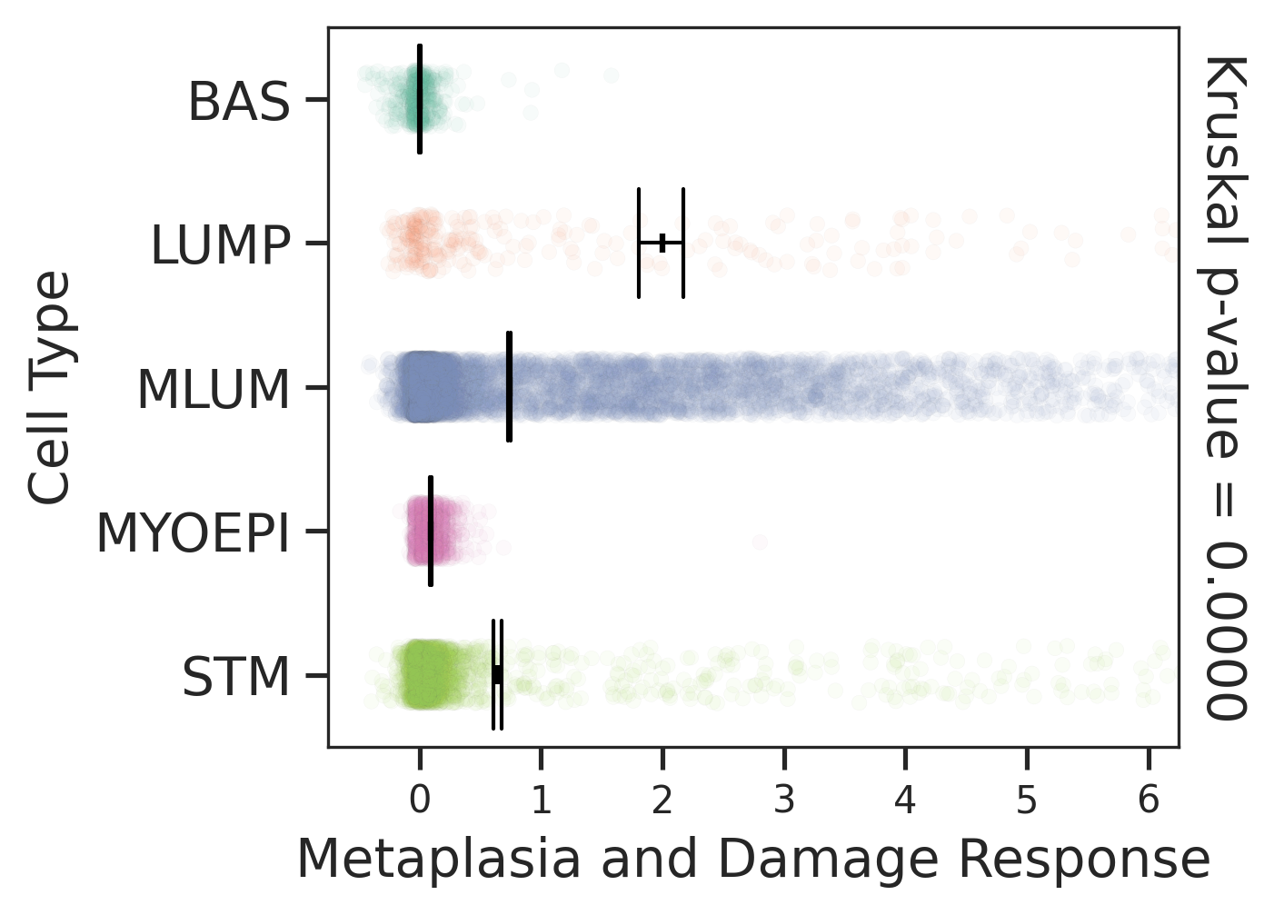
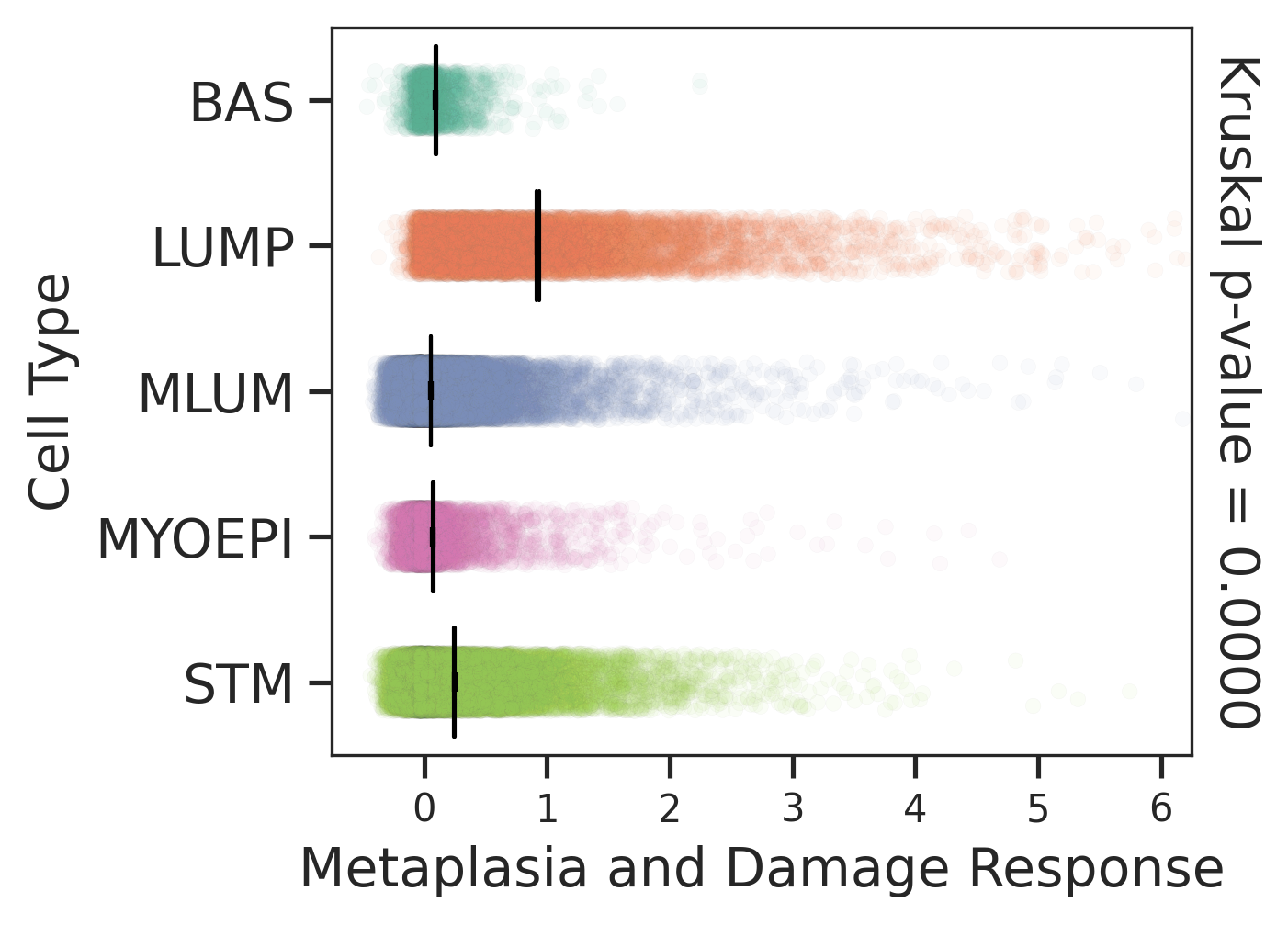
check button Metaplasia and damage response signature (for epithelial cells).
HealthyPrecancerDCISIDC
Comparisons of Metaplasia-associated damage scores among epithelial cell subpopulations.Comparisons of Metaplasia-associated damage scores among epithelial cell subpopulations.Comparisons of Metaplasia-associated damage scores among epithelial cell subpopulations.Comparisons of Metaplasia-associated damage scores among epithelial cell subpopulations.
∗Comparisons of Metaplasia-associated damage scores among epithelial cell subpopulations.

check button Neoantigen-specific TCR clonotypes (NeoTCR) signature.
Comparisons of CD4+ and CD8+ NeoTCR signature scores among different disease states.Comparisons of CD4+ and CD8+ NeoTCR signature scores among different disease states.
∗Comparisons of CD4+ and CD8+ NeoTCR signature scores among different disease states.